
Stock image Funduscopic
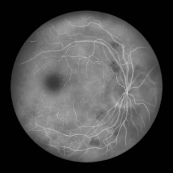
Non-proliferative Diabetic Retinopathy, Illustration Showing Cotton Wool Spots As Fluffy Dark Patches, Abnormal Finding On Funduscopic Examination Of The Eye Retina In Diabetes Mellitus, Fluorescein Angiography
Image, 2.72MB, 5000 × 5000 jpg

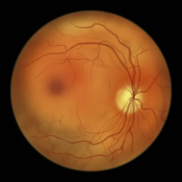
Non-proliferative Diabetic Retinopathy, 3D Illustration Showing Normal Eye Retina And Retina With Hard Exudates (irregularly Shaped Yellow Spots)
Image, 25.27MB, 11738 × 6603 jpg

Non-proliferative Diabetic Retinopathy, Illustration Showing Normal Eye Retina And Retina With Hard Exudates, Microaneurysms, Dot Haemorrhages, Flame-shaped And Splinter Retinal Haemorrhages
Image, 6.89MB, 11738 × 6603 jpg
Non-proliferative Diabetic Retinopathy, Illustration Showing IRMAs (intraretinal Microvascular Abnormalities) As Small Vessels With Abnormal Branching Or Dilatation In Ischaemic Areas
Image, 2.68MB, 5000 × 5000 jpg
Normal Eye Retina, Ophthalmoscope View, Scientific 3D Illustration Showing Optic Disk, Blood Vessels, Macula And Fovea
Image, 13.02MB, 5352 × 5352 jpg

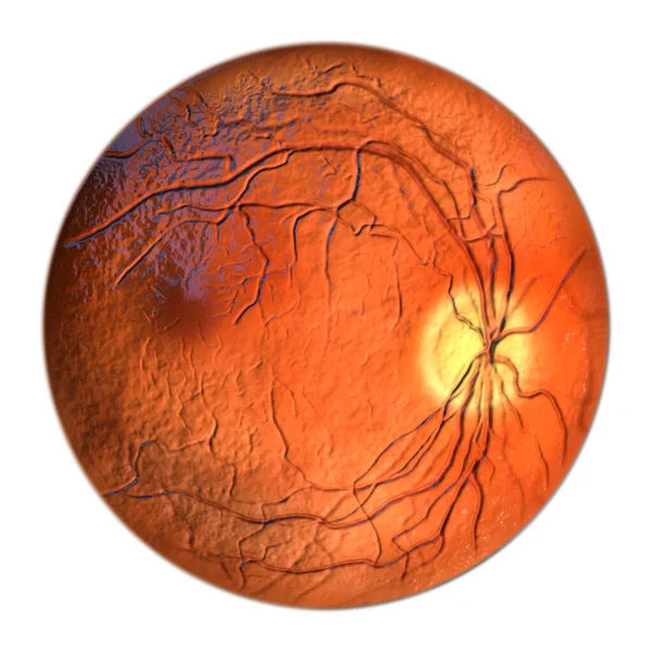
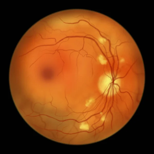
Non-proliferative Diabetic Retinopathy, 3D Illustration Showing Hard Exudates, Microaneurysms, Dot Haemorrhages, Flame-shaped And Splinter Retinal Haemorrhages, Ophthalmoscope View
Image, 13.11MB, 5352 × 5352 jpg
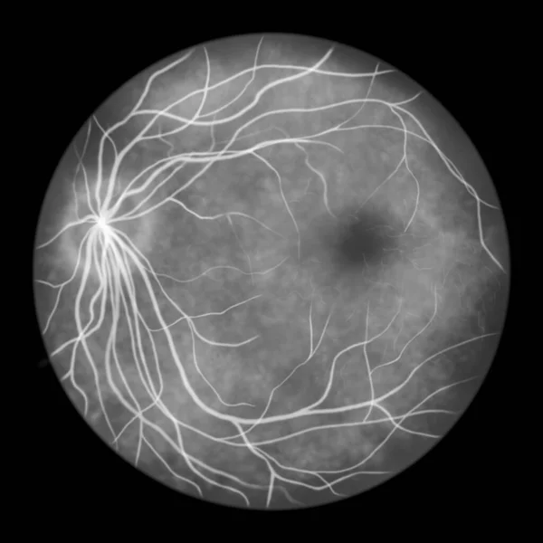
Normal Eye Retina, Scientific Illustration Showing Optic Disk, Blood Vessels, Macula And Fovea, Ophthalmoscope View, Fluorescein Angiography
Image, 2.91MB, 5000 × 5000 jpg

Non-proliferative Diabetic Retinopathy, 3D Illustration Showing Normal Eye Retina And Retina With Hard Exudates, And Cotton Wool Spots
Image, 25.26MB, 11738 × 6603 jpg

Normal Eye Retina, Ophthalmoscope View, Scientific Illustration Showing Optic Disk, Blood Vessels, Macula And Fovea
Image, 2.74MB, 5000 × 5000 jpg
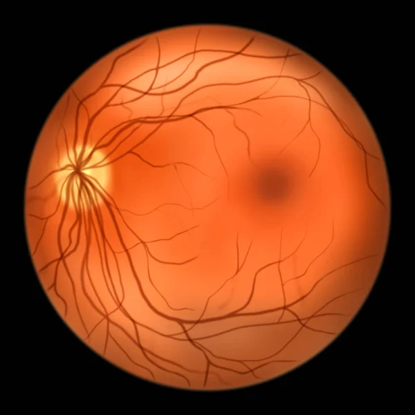
Normal Eye Retina, Ophthalmoscope View, Scientific Illustration Showing Optic Disk, Blood Vessels, Macula And Fovea
Image, 2.88MB, 5000 × 5000 jpg

Non-proliferative Diabetic Retinopathy, Illustration Showing Normal Eye Retina And Retina With Cotton Wool Spots As Fluffy Yellow Patches
Image, 6.22MB, 11738 × 6603 jpg

Non-proliferative Diabetic Retinopathy, Illustration Showing Normal Eye Retina And Retina With Hard Exudates, Microaneurysms, Dot Haemorrhages, Flame-shaped And Splinter Retinal Haemorrhages
Image, 7.35MB, 11738 × 6603 jpg
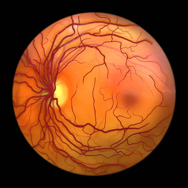
Normal Eye Retina, Ophthalmoscope View, Scientific 3D Illustration Showing Optic Disk, Blood Vessels, Macula And Fovea
Image, 11.53MB, 5000 × 5000 jpg

Non-proliferative Diabetic Retinopathy, Illustration Showing IRMAs (intraretinal Microvascular Abnormalities), Venous Beading, And Microaneurysms
Image, 2.85MB, 5000 × 5000 jpg
Non-proliferative Diabetic Retinopathy, Illustration Showing Cotton Wool Spots As Fluffy Yellow Patches, Abnormal Finding On Funduscopic Examination Of The Eye Retina In Diabetes Mellitus
Image, 2.7MB, 5000 × 5000 jpg
Page 1 >> Next