Home
Images
Vectors
Videos
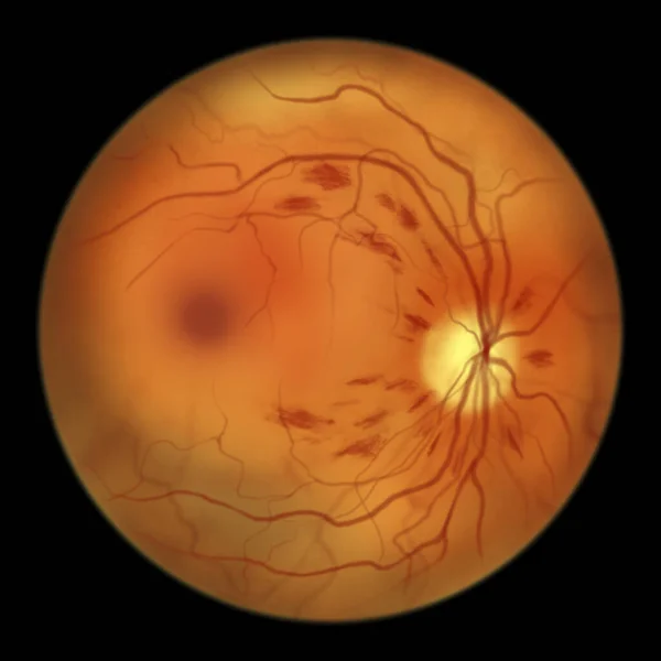
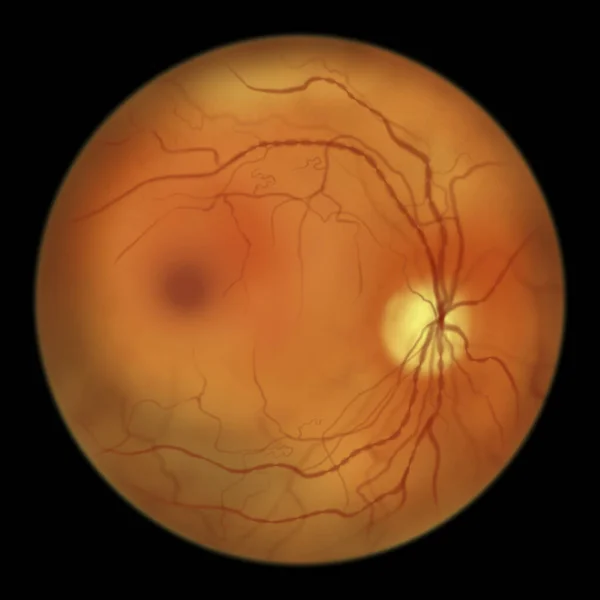
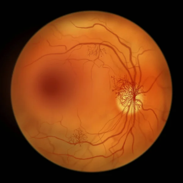
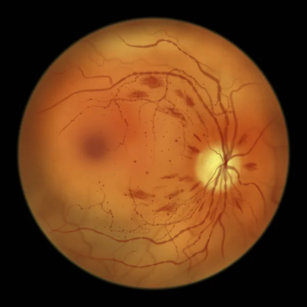
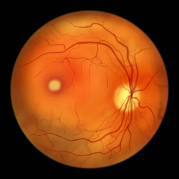
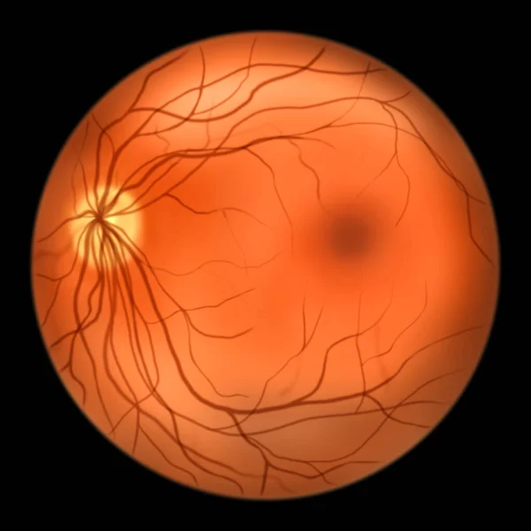
Stock image Normal eye retina, ophthalmoscope view, scientific illustration showing optic disk, blood vessels, macula and fovea
Published:
Apr.14, 2022 07:11:25
Author:
katerynakon
Views:
27
Downloads:
2
File type:
image
/
jpg
File size:
2.88 MB
Orginal size:
5000
x
5000
px
Available sizes:
S
(500 x 500)
M
(1000 x 1000)
L
(2000 x 2000)
XL
(5000 x 5000)
EL0
(5000 x 5000)
Level:
silver
Download Image
Image topics:
Image
Illustration
Health
Healthy
Blood
Pattern
Medicine
Illness
Medical
Black
Eyes
Doctor
Hospital
Exam
Patient
Vessel
Vision
Organ
Disk
Vein
Optic
Membrane
Examination
Physician
Normal
Investigation
Ophthalmologist
Ocular
Ophthalmology
Artery
Diseases
Retina
Nerve
Ophthalmoscopy
Retinal
Fovea
Fundus
Macula
Health Care
Hand Drawn
Hand Drawing
Retinoscopy
Left Eye
Optic Disk
Neuroretinal
Funduscopic
Optic Cup
Similar stock images
Non-proliferative Diabetic Retinopathy, Illustration Showing Flame-shaped And Splinter Retinal Haemorrhages, Ophthalmoscope View
5000 × 5000
Non-proliferative Diabetic Retinopathy, Illustration Showing IRMAs (intraretinal Microvascular Abnormalities) As Small Vessels With Abnormal Branching In Ischaemic Areas, And Venous Beading
5000 × 5000
Proliferative Diabetic Retinopathy, Illustration Showing Neovascularization In The Disk And Other Sites, And Macula Edema. Fundoscopic Examination Of The Eye Retina In Diabetes Mellitus
5000 × 5000
Normal Eye Retina, Ophthalmoscope View, Scientific Illustration Showing Optic Disk, Blood Vessels, Macula And Fovea
5000 × 5000
Non-proliferative Diabetic Retinopathy, Illustration Showing IRMAs (intraretinal Microvascular Abnormalities), Microaneurysms, Dot Haemorrhages, Flame-shaped And Splinter Retinal Haemorrhages
5000 × 5000
Non-proliferative Diabetic Retinopathy, Illustration Showing Small Retinal Haemorrhages, Ophthalmoscope View
5000 × 5000
Best Disease. Best Vitelliform Macular Dystrophy, Vitelliform Stage, Classic Egg-yolk Lesion, Scientific Illustration, Ophthalmoscope View
5000 × 5000