
Stock image Mhc
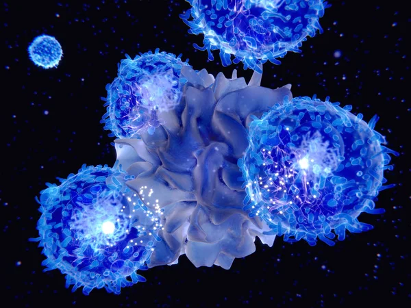
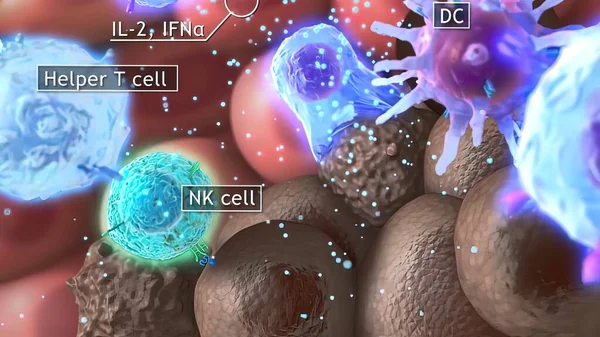
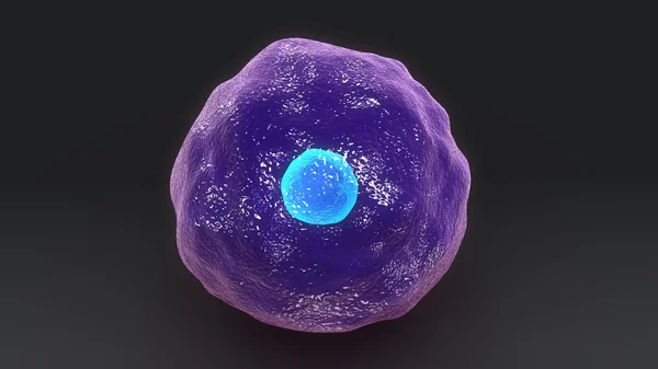
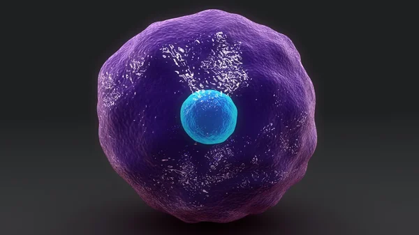
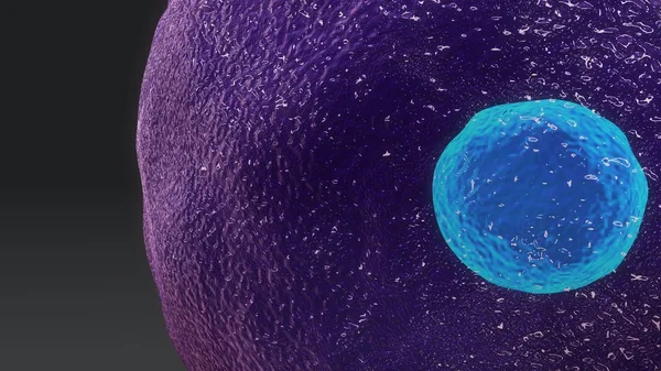
3d Computer Illustration Of A Dendritic Cell. They Areantigen-presenting Cells Of The Immune System. Their Main Function Is To Process Antigen Material And Present It On The Cell Surface To The T Cells Of The Immune System. They Are Messengers Betwe
Image, 5.2MB, 8000 × 6000 jpg
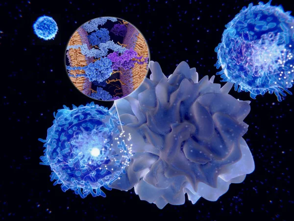
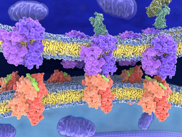
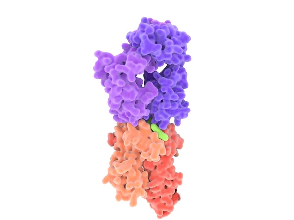
Dendritic Cells Present Antigens (green) To Lymphocytes Through Their Membran Bound MHC-molecules (violet). CD4 Molecules (light Blue) Bind To Other Portions Of The MHC, Strengthening The Interaction.
Image, 10.24MB, 8000 × 6000 jpg
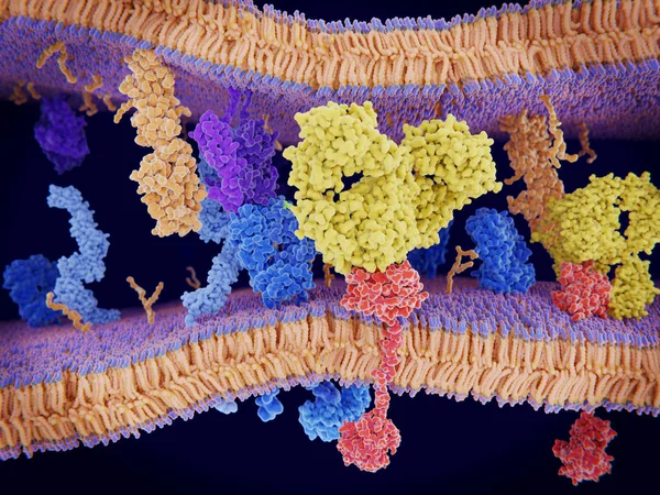
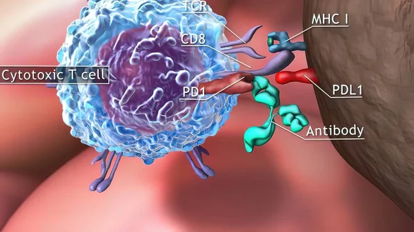
Cancer Cells Express PD-L1 (orange) Proteins On Their Surface To Trick The Immune System. The Interaction Of PD-L1 With PD-1 Of T-cells Leads To A Down-regulation Of T-cells. The Antibody (yellow) Blocks This Interaction.
Image, 18.3MB, 8000 × 6000 jpg
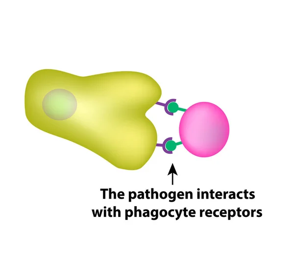
Innate Immunity. Adaptive Specific . Phagocytosis. Infographics. Vector Illustration
Vector, 1.45MB, 5000 × 4734 eps
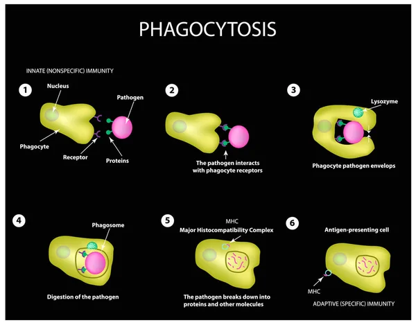
Innate Immunity. Adaptive Specific . Phagocytosis. Infographics. Vector
Vector, 3.99MB, 5000 × 3911 eps

BARCELONA, SPAIN - MARCH 1, 2022: The Photography Section Within The Museum D'Historia De Catalunya Located In The Building Palau De Mar (1880-1890), Originally General Port Warehouse, A 19th-century.
Image, 8.71MB, 5315 × 3544 jpg
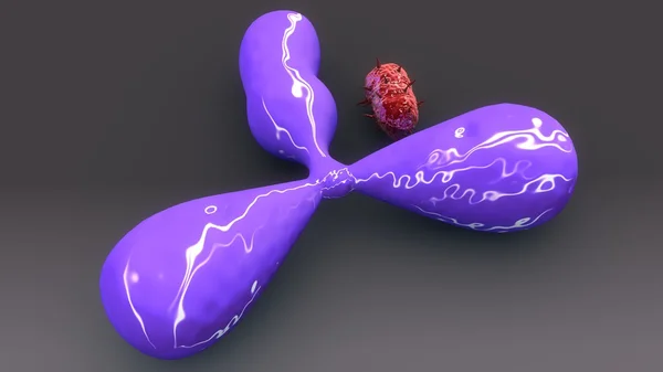
3D Medical Illustration Cytotoxic T Cell And Helper T Cell Destroys The Stress-related Molecule
Image, 1.77MB, 3840 × 2160 jpg
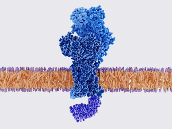
The T-cell Receptor Activates The Immune Response To Antigens In T-lymphocytes. T-cell Receptors (dark Blue), CD4 Molecules (light Blue), Glycolipids (orange). 3d Rendering. Illustration
Image, 3.11MB, 8000 × 6000 jpg
T-cell Receptor In Complex With The MHC Class II-peptide Complex. The Antigen (light Green) Is A Peptide From A Tumor Cell, Bacteria Or Virus. Different Stages Of The Interaction. 3D-Rendering. Illustration
Image, 9.21MB, 8000 × 6000 jpg

Vector Illustration Of Mechanisms Of Microbial Cell Resistance To Antibiotics
Vector, 3.53MB, 6688 × 3296 eps


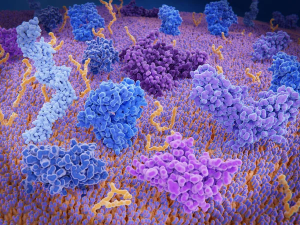
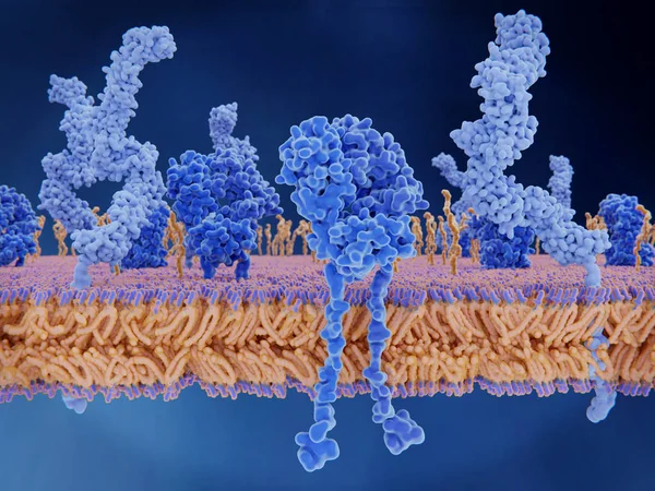
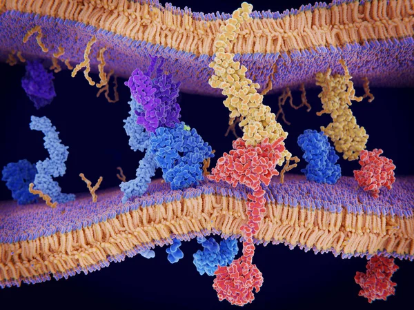
Immunologically Active Proteins On A T-cell. TCR (blue), CD-4 (light Blue), CD-28 (dark Blue), PD-1 (magenta), CTLA-4 (violet), Ca-channel (dark Violet). The T-cell Receptor, CD-4 And CD-28 Activate T-cells, While PD-1 And CTLA-4 Inhibit The Activat
Image, 10.2MB, 8000 × 6000 jpg

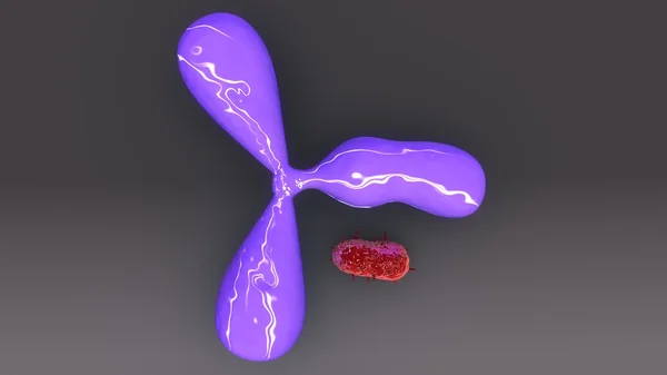
Macrophages Approaching Bacteria (bacilli), 3D Rendering. Illustration
Image, 2.49MB, 8000 × 6000 jpg
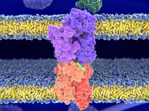
T-cell Receptor In Complex With The MHC Class II-peptide Complex. The Antigen (light Green) Is A Peptide From A Tumor Cell, Bacteria Or Virus. Complex Embedded In The Membranes. 3D-Rendering. Illustration
Image, 7.59MB, 8000 × 6000 jpg

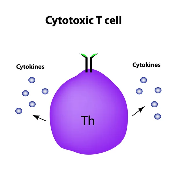
Cytotoxic Cells. Cytokines. Cell Immunity. Infographics. Vector Illustration
Vector, 1.39MB, 5000 × 5000 eps

T-cell Receptor In Complex With The MHC Class II-peptide Complex. The Antigen (light Green) Is A Peptide From A Tumor Cell, Bacteria Or Virus. Different Stages Of The Interaction. 3D-Rendering. Illustration
Image, 7.31MB, 8000 × 6000 jpg
MHC Letter Logo Design For Technology Company. MHC Logo Design Black And White Color Combination. MHC Logo, MHC Vector, MHC Design, MHC Icon, MHC Alphabet. MHC Typography Logo Design.
Vector, 5.02MB, 9792 × 9793 eps

Innate Immunity. Adaptive Specific . Phagocytosis. Infographics. Vector Illustration
Vector, 1.48MB, 5000 × 4734 eps

Lymphocyte B And T-cell. Cells Of Immune System (immune Response). Vector Illustration On White Background. Didatic Illustration.
Vector, 0.49MB, 5000 × 5000 ai

Vector Illustration Of Mechanisms Of Microbial Cell Resistance To Antibiotics
Vector, 3.53MB, 6688 × 3296 eps
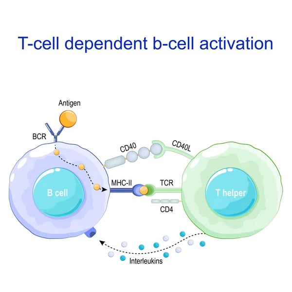
T-cell Dependent B-cell Activation. B Lymphocyte Binds An Antigen, Receive Help From A T Helper, And Differentiate Into A Plasma Cell That Secretes Of Antibodies. Receptors On Surface Of White Blood Cells. Human Immune System. Vector Poster
Vector, 1.05MB, 4444 × 4444 eps
T-cell Receptor In Complex With The MHC Class II-peptide Complex. The Antigen (light Green) Is A Peptide From A Tumor Cell, Bacteria Or Virus. Different Stages Of The Interaction. 3D-Rendering. Illustration
Image, 2.17MB, 8000 × 6000 jpg

BARCELONA, SPAIN - MARCH 1, 2022: Museu D'Historia De Catalunya Located In The Building Palau De Mar (1880-1890), Originally General Port Warehouse, A 19th-century Warehouse In Front Of The Harbor.
Image, 14.15MB, 5315 × 3543 jpg
PD-1 (red) Extends From The Surface Of A T-cell And Interacts With The Ligand Protein PD-L1 (yellow) From A Antigen Presenting Cell. Although The T-cell Has Been Activated Through The Interaction Of A T-cell Receptor (blue) And A MHC Protein (viole
Image, 18.32MB, 8000 × 6000 jpg

3D Medical Illustration Cytotoxic T Cell And Helper T Cell Destroys The Stress-related Molecule
Image, 1.44MB, 3840 × 2160 jpg
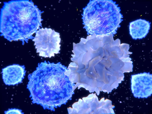
T-lymphocytes And Dendritic Cells, 3D-rendering; Dendritic Cells Are Antigen-presenting Cells Of The Immune System. They Process Antigen Material And Present It On The Cell Surface To The T-cells. Illustration
Image, 2.23MB, 4000 × 3000 jpg
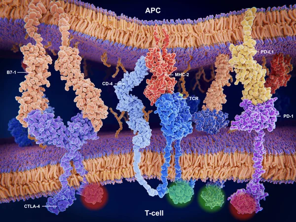
Interactions Of MHC-II With The T-cell Receptor And CD4 And B7-1 With CD-28 Activates T-cells While The Interactions Of P7-1 With CTLA-4 And PD-L1 With PD-1 Deactivates T-cells.
Image, 10.7MB, 8000 × 6000 jpg
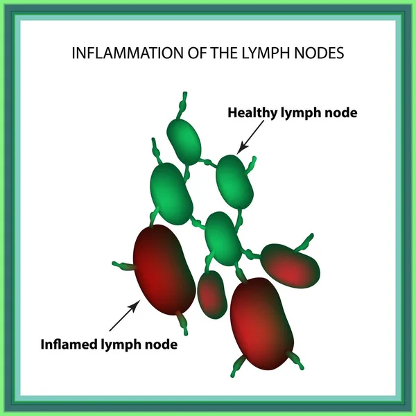
Inflammation Of The Lymph Nodes. Infographics. Vector Illustration On Isolated Background
Vector, 1.13MB, 5000 × 5000 eps
Macrophag Engulfing Bacteria (cocci), 3D Rendering. Macrophages Engulf And Digest Cellular Debris And Pathogens. - Illustration
Image, 2.72MB, 8000 × 6000 jpg

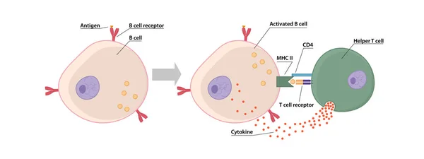
T And B Cell Binding To Elicit A Response To A T Cell-dependent Antigen, The B And T Cells Must Come Close Together. B Cell Must Receive Two Signals From The Native Antigen And The T Cells Cytokines
Vector, 5.86MB, 8334 × 3125 eps
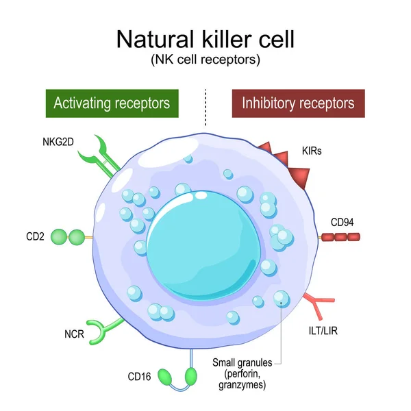
Natural Killer Cell. NK Cell Receptors. Structure And Anatomy Of Large Granular Lymphocytes (LGL). Human Immune System. Part Of Innate Immunity. Vector Poster
Vector, 1.22MB, 4444 × 4444 eps
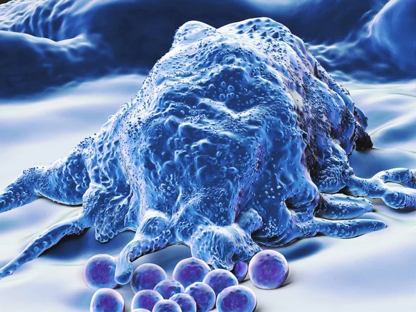
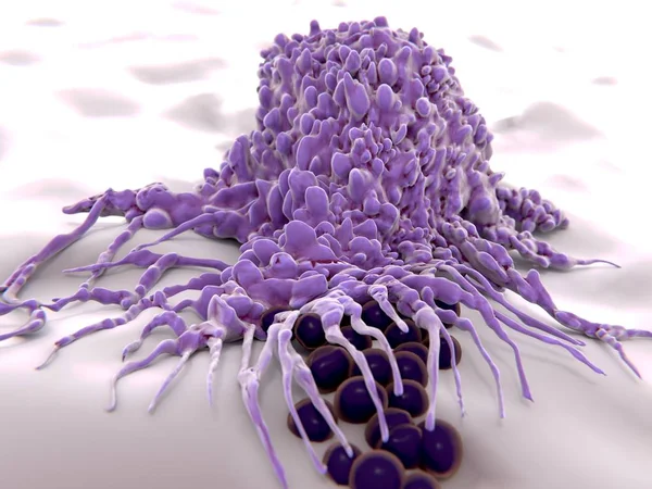
Makrophage, Type Of White Blood Cell, Of The Immune System, That Engulfs And Digests Cellular Debris, Foreign Substances, Microbes, Cancer Cells
Image, 9.6MB, 4000 × 3000 jpg

Activation Of The Immune Response To An Antigene (green) Through The Complex Between A T-cell Receptor (dark Blue), An MHC II-antigen (violet) And A CD4 Protein (light Blue). 3d Rendering. Illustration
Image, 6.36MB, 8000 × 6000 jpg
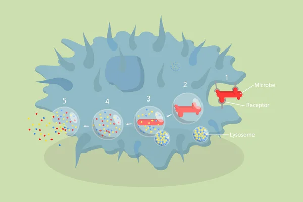
3D Isometric Flat Vector Conceptual Illustration Of Phagocytosis, Labeled Endocytosis Educational Scheme
Vector, 1.75MB, 6000 × 4000 eps
Page 1 >> Next