
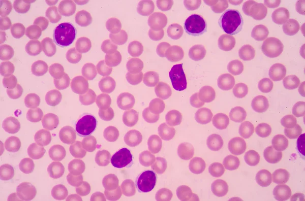
Stock image Mhc Complex
The T-cell Receptor Activates The Immune Response To Antigens In T-lymphocytes. T-cell Receptors (dark Blue), CD4 Molecules (light Blue), Glycolipids (orange). 3d Rendering. Illustration
Image, 3.11MB, 8000 × 6000 jpg

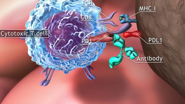
Cancer Cells Express PD-L1 (orange) Proteins On Their Surface To Trick The Immune System. The Interaction Of PD-L1 With PD-1 Of T-cells Leads To A Down-regulation Of T-cells. The Antibody (yellow) Blocks This Interaction.
Image, 18.3MB, 8000 × 6000 jpg
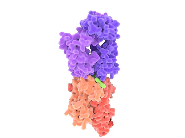
T-cell Receptor In Complex With The MHC Class II-peptide Complex. The Antigen (light Green) Is A Peptide From A Tumor Cell, Bacteria Or Virus. Complex Embedded In The Membranes. 3D-Rendering. Illustration
Image, 7.59MB, 8000 × 6000 jpg

Interactions Of MHC-II With The T-cell Receptor And CD4 And B7-1 With CD-28 Activates T-cells While The Interactions Of P7-1 With CTLA-4 And PD-L1 With PD-1 Deactivates T-cells.
Image, 10.7MB, 8000 × 6000 jpg

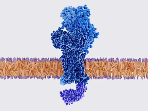
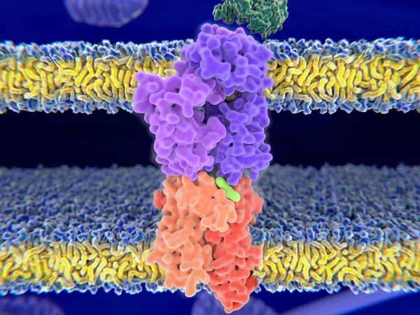
Activation Of The Immune Response To An Antigene (green) Through The Complex Between A T-cell Receptor (dark Blue), An MHC II-antigen (violet) And A CD4 Protein (light Blue). 3d Rendering. Illustration
Image, 6.36MB, 8000 × 6000 jpg
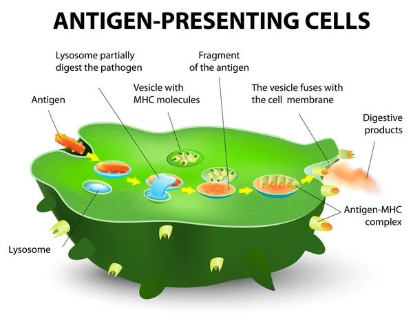
Dendritic Cell. Dendritic Cells Are Antigen-presenting Cells Of The Immune System. They Process Antigen Material And Present It On The Cell Surface To The T Cells Of The Immune System. 3D Rendering. Illustration
Image, 2.94MB, 8000 × 6000 jpg


T-cell Receptor In Complex With The MHC Class II-peptide Complex. The Antigen (light Green) Is A Peptide From A Tumor Cell, Bacteria Or Virus. Different Stages Of The Interaction. 3D-Rendering. Illustration
Image, 7.31MB, 8000 × 6000 jpg
PD-1 (red) Extends From The Surface Of A T-cell And Interacts With The Ligand Protein PD-L1 (yellow) From A Antigen Presenting Cell. Although The T-cell Has Been Activated Through The Interaction Of A T-cell Receptor (blue) And A MHC Protein (viole
Image, 18.32MB, 8000 × 6000 jpg
T-cell Receptor In Complex With The MHC Class II-peptide Complex. The Antigen (light Green) Is A Peptide From A Tumor Cell, Bacteria Or Virus. Different Stages Of The Interaction. 3D-Rendering. Illustration
Image, 2.17MB, 8000 × 6000 jpg
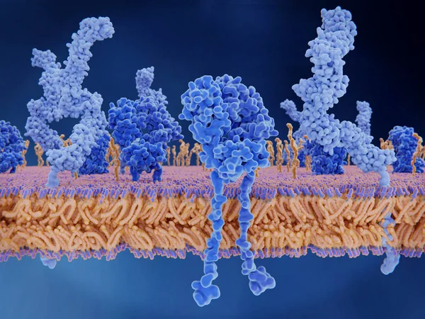
Dendritic Cells Present Antigens (green) To Lymphocytes Through Their Membran Bound MHC-molecules (violet). CD4 Molecules (light Blue) Bind To Other Portions Of The MHC, Strengthening The Interaction.
Image, 10.24MB, 8000 × 6000 jpg
T-cell Receptor In Complex With The MHC Class II-peptide Complex. The Antigen (light Green) Is A Peptide From A Tumor Cell, Bacteria Or Virus. Different Stages Of The Interaction. 3D-Rendering. Illustration
Image, 9.21MB, 8000 × 6000 jpg

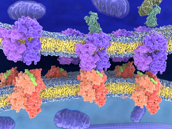
Interaction Of MHC-II (red) With The T-cell Receptor (blue) And CD4 (light Blue) And B7-1 (orange) With CD-28 (dark Blue) Activates T-cells While The Interaction Of P7-1 With CTLA-4 (violett) And PD-L1 (yellow) With PD-1 Deactivates T-cells
Image, 10.65MB, 8000 × 6000 jpg
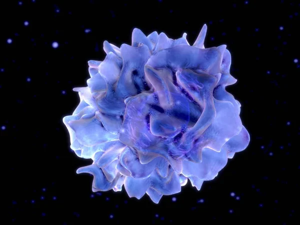
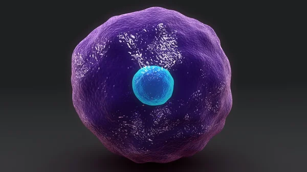
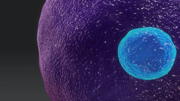
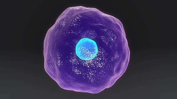
Dendritic Cell. 3D-rendering. Dendritic Cells Are Antigen-presenting Cells Of The Immune System. Illustration
Image, 5.07MB, 8000 × 6000 jpg
3d Computer Illustration Of A Dendritic Cell. They Areantigen-presenting Cells Of The Immune System. Their Main Function Is To Process Antigen Material And Present It On The Cell Surface To The T Cells Of The Immune System. They Are Messengers Betwe
Image, 5.2MB, 8000 × 6000 jpg
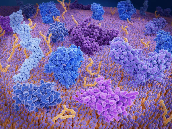
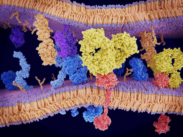
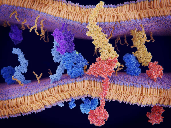
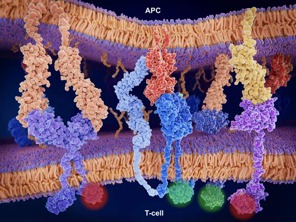
Immunologically Active Proteins On A T-cell. TCR (blue), CD-4 (light Blue), CD-28 (dark Blue), PD-1 (magenta), CTLA-4 (violet), Ca-channel (dark Violet). The T-cell Receptor, CD-4 And CD-28 Activate T-cells, While PD-1 And CTLA-4 Inhibit The Activat
Image, 10.2MB, 8000 × 6000 jpg
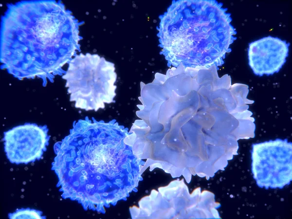
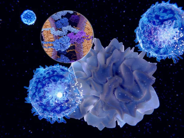
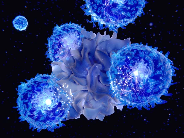
T-lymphocytes And Dendritic Cells, 3D-rendering; Dendritic Cells Are Antigen-presenting Cells Of The Immune System. They Process Antigen Material And Present It On The Cell Surface To The T-cells. Illustration
Image, 2.23MB, 4000 × 3000 jpg
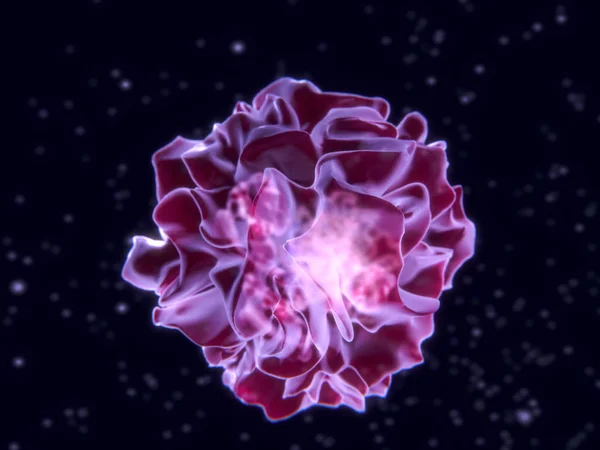
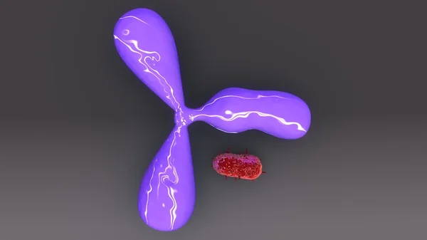
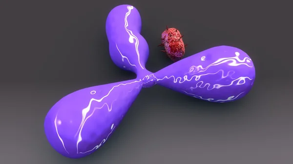
3D Medical Illustration Cytotoxic T Cell And Helper T Cell Destroys The Stress-related Molecule
Image, 1.77MB, 3840 × 2160 jpg
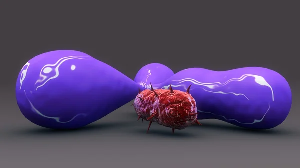
3D Medical Illustration Cytotoxic T Cell And Helper T Cell Destroys The Stress-related Molecule
Image, 1.44MB, 3840 × 2160 jpg


3D Medical Illustration Cytotoxic T Cell And Helper T Cell Destroys The Stress-related Molecule
Image, 1.78MB, 3840 × 2160 jpg
Page 1 >> Next