
Stock image Micrography
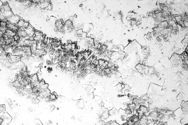


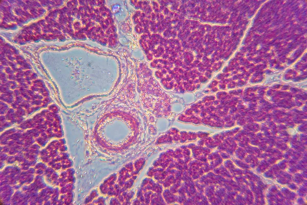
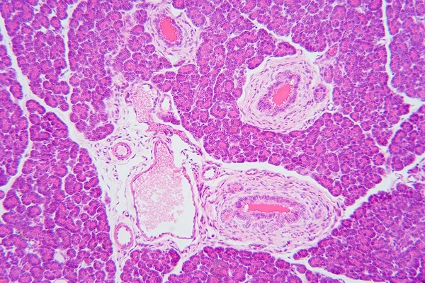
This Is A Histological Photograph Of The Human Small Intestine. Magnify 40x.
Image, 9.15MB, 4202 × 4202 jpg


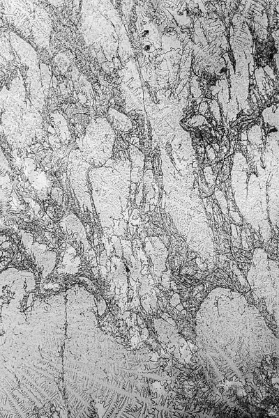
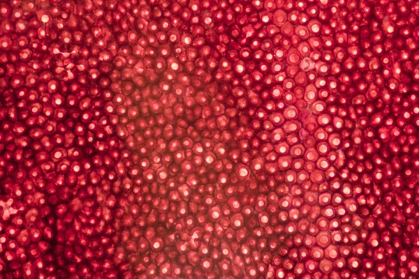
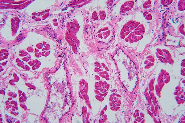
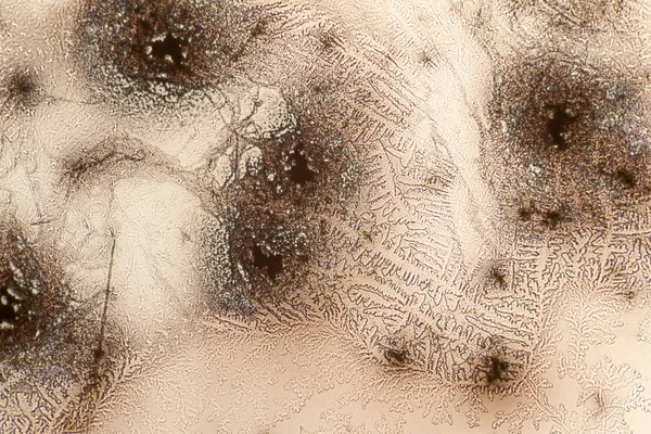
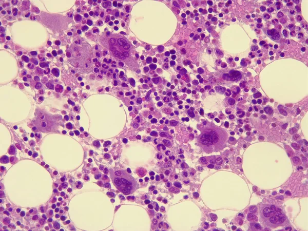
This Is A Pathological Photo Of Human Left Ventricular Hypertrophy, Showing An Increase In Myocardial Diameter And Interstitial Distance.Magnify 40x.
Image, 42.86MB, 8500 × 8500 jpg

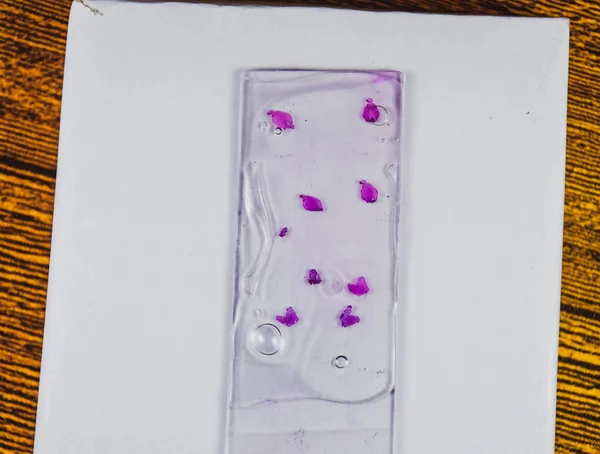
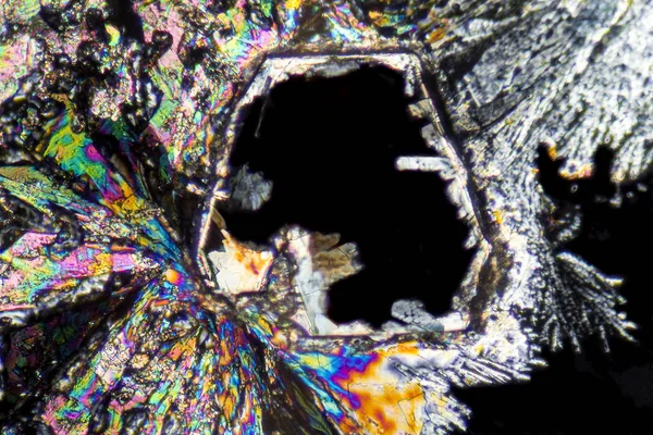
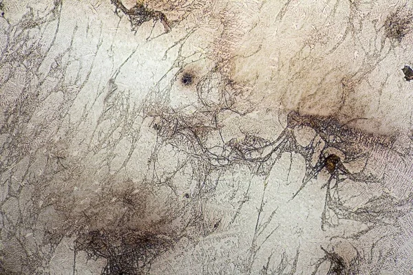
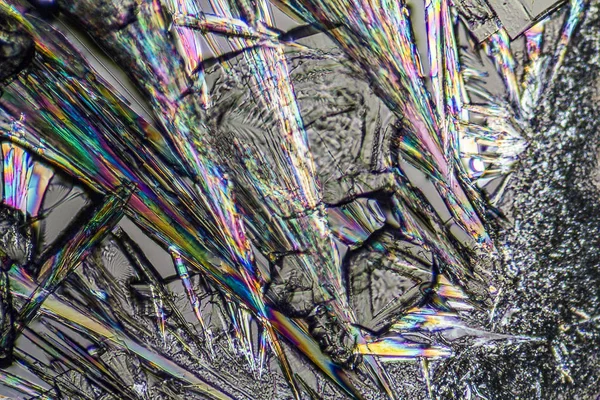
Slices Of The Tumor Under Glass. Histological Examination Of Tumor Cells For The Presence Of Cancer
Image, 1.41MB, 2475 × 1873 jpg


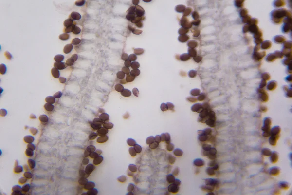
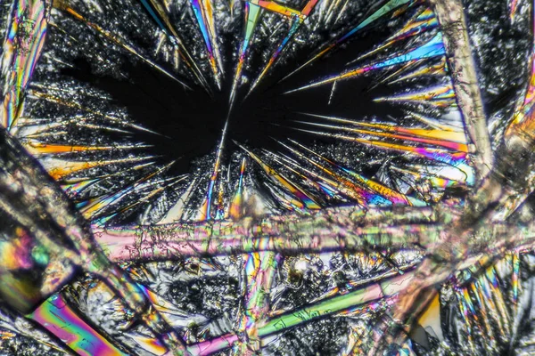
This Photo Shows The Pink Mucinous Stroma Of An Atrial Myxoma And The Myxoma Cells Arranged In A Nested And Cord-like Pattern.Magnify 1000x.
Image, 42.93MB, 7000 × 7000 jpg


This Photo Shows The Pink Mucinous Matrix And Nest Like Arrangement Of Mucinous Tumor Cells In Atrial Myxoma.Magnify 1000x.
Image, 42.8MB, 6700 × 6700 jpg

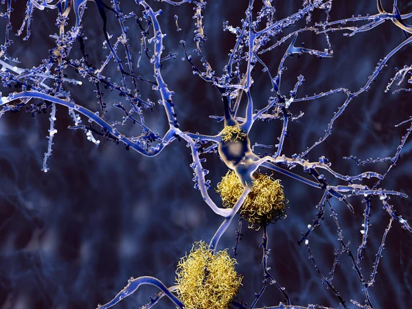
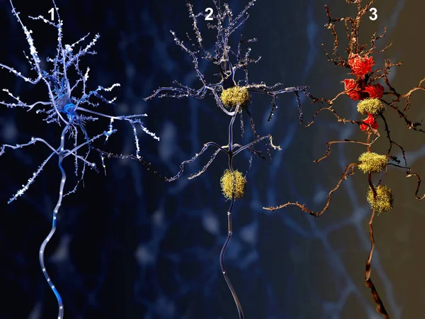
3 Phases Of The Alzheimer Disease. 1. Healthy Neuron. 2. Neuron With Amyloid Plaques (yellow). 3. Dead Neuron Being Digested By Microglia Cells (red). Illustration
Image, 6.76MB, 8000 × 6000 jpg

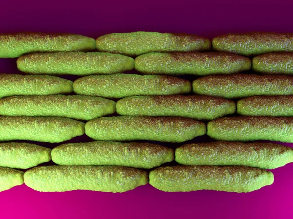
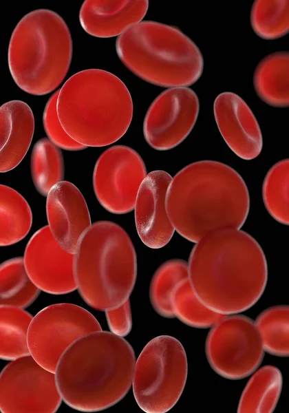
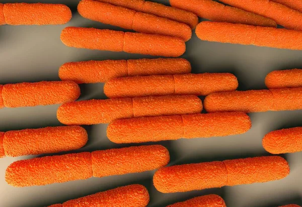
Cancer Cells Mitosis Or Proliferation 3D Rendering Illustration. Division Of Two Malignant Cells Causing Carcinoma Close-up. Medicine, Oncology, Science, Disease, Biology And Microbiology Concepts.
Image, 4.56MB, 3840 × 2160 jpg

Photomicrograph Showing The Dorsal View (from Above) Of A Live Microscopic Water Bear (tardigrade)
Image, 8.05MB, 3645 × 2734 jpg

This Is A Histological Photograph Of The Human Small Intestine. Magnification 40x
Image, 41.82MB, 8000 × 8000 jpg
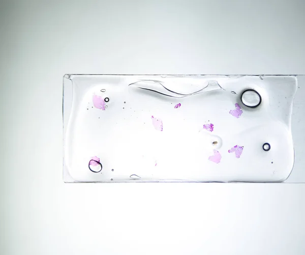
Slices Of The Tumor Under Glass. Histological Examination Of Tumor Cells For The Presence Of Cancer
Image, 1.3MB, 2800 × 2340 jpg


3 Phases Of The Alzheimer Disease. 1. Healthy Neuron. 2. Neuron With Amyloid Plaques (yellow). 3. Dead Neuron Being Digested By Microglia Cells (red). Illustration
Image, 6.76MB, 8000 × 6000 jpg
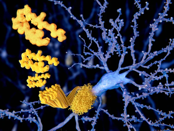
The Beta Amyloid Peptid, Amyloid Plaques Growing On A Neuron. It Consists Of About 30 Amino Acids And Aggregates To Amyloid Plaques, That May Damage And Kill Neurons. Illustration
Image, 4.49MB, 8000 × 6000 jpg
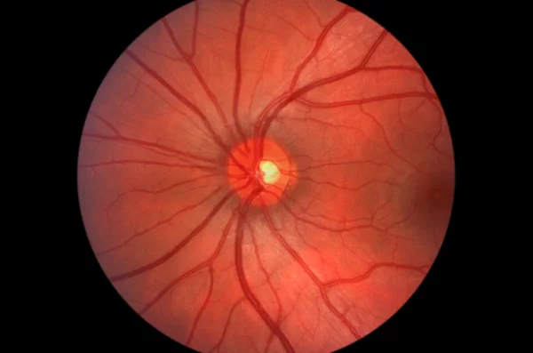


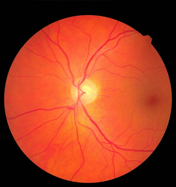
Ophthalmic Image Detailing The Retina And Optic Nerve Inside A Healthy Human Eye. Medicine Concept
Image, 3.41MB, 3000 × 3186 jpg

3/4 View Photomicrograph Of A Tiny Springtail (Collembola) Showing Its Internal Anatomy
Image, 5.92MB, 5222 × 3903 jpg

Slices Of The Tumor Under Glass. Histological Examination Of Tumor Cells For The Presence Of Cancer
Image, 2.65MB, 4000 × 3484 jpg


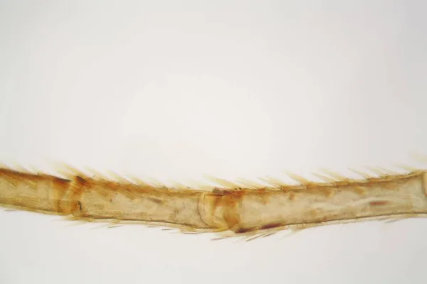
Photomicrograph Of The Side View Of A Minute Springtail (Collembola) Showing Its Internal Anatomy
Image, 5.57MB, 5449 × 4071 jpg

Page 1 >> Next