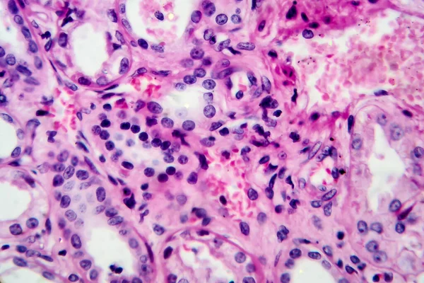
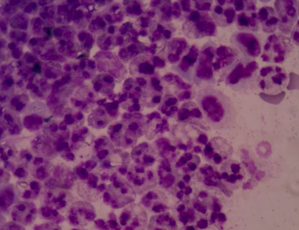
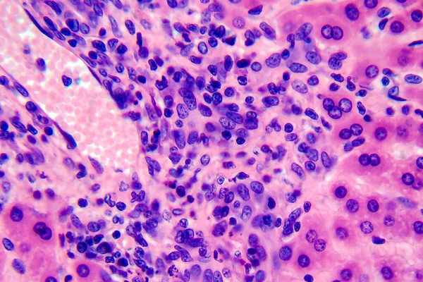
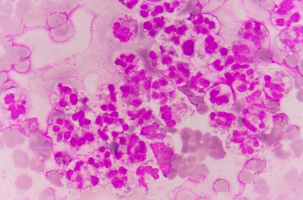
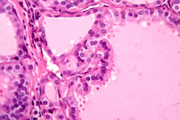
Stock image This photo shows the pink mucinous matrix and nest like arrangement of mucinous tumor cells in atrial myxoma.Magnify 1000x.

Published: May.11, 2024 12:24:31
Author: asia11m
Views: 0
Downloads: 0
File type: image / jpg
File size: 42.8 MB
Orginal size: 6700 x 6700 px
Available sizes:
Level: beginner