
Stock image Ophthalmic Disorders
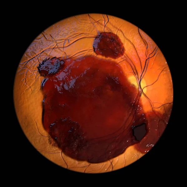
Medical 3D Illustration Of A Subretinal Hemorrhage Observed During Ophthalmoscopy, Revealing A Dark, Irregular Hemorrhage Beneath The Retinal Layers.
Image, 9.37MB, 5352 × 5352 jpg

A Prepapillary Vascular Loop On The Retina, As Observed During Ophthalmoscopy In Fluorescein Angiogram, An Illustration Showcasing The Looping Blood Vessels Around The Optic Disc.
Image, 2.77MB, 5000 × 5000 jpg
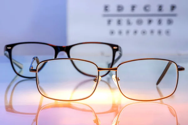
Eyeglasses With Prescription Lenses On A Backlit Glass Table Against A Visual Test With Letters In Blur Background. Vision Correction And Ophthalmology Concept. Selected Focus
Image, 10.64MB, 6773 × 4521 jpg

Retina In Blastomycosis (infection Caused By Fungi Blastomyces Dermatitidis) As Seen During Ophthalmoscopy. An Illustration Showing Scattered Yellow Choroidal Infiltrates And A Choroidal Mass Lesion.
Image, 2.86MB, 5000 × 5000 jpg

A Prepapillary Vascular Loop On The Retina, As Observed During Ophthalmoscopy, An Illustration Showcasing The Looping Blood Vessels Around The Optic Disc.
Image, 7.68MB, 11738 × 6603 jpg

A 3D Illustration Of Valsalva Retinopathy Observed During Ophthalmoscopy, Showcasing Retinal Hemorrhages Resulting From Sudden Increase In Intraocular Pressure With Characteristic Double Ring Sign.
Image, 20.34MB, 11738 × 6603 jpg

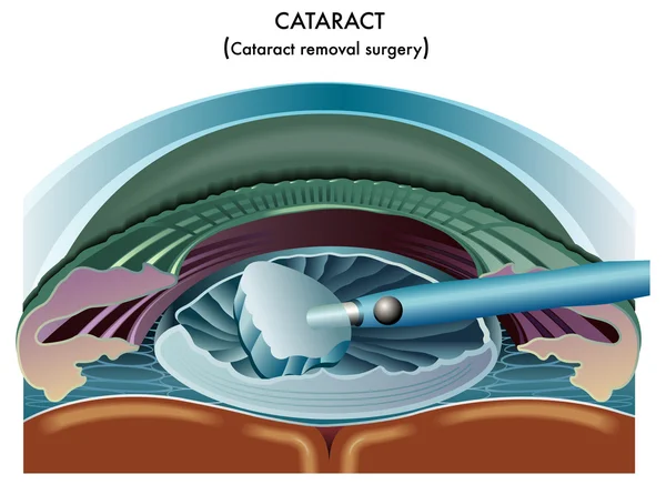
The Use Of Medical Lasers To Cutting Eye Lens In Small Pieces And Suction Out, In Order To Use Artificial Lenses, Because The Symptoms Are Cataracts.
Vector, 10.65MB, 6000 × 6000 eps

Medical 3D Illustration Of A Subretinal Hemorrhage Observed During Ophthalmoscopy, Revealing A Dark, Irregular Hemorrhage Beneath The Retinal Layers.
Image, 9.71MB, 5352 × 5352 jpg

A Medical Illustration Depicting Vitreous Hemorrhage Observed During Ophthalmoscopy, Revealing Blood Within The Vitreous Humor Obscuring Retinal Details.
Image, 2.99MB, 5000 × 5000 jpg
Page 1 >> Next