Home
Images
Vectors
Videos
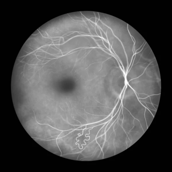
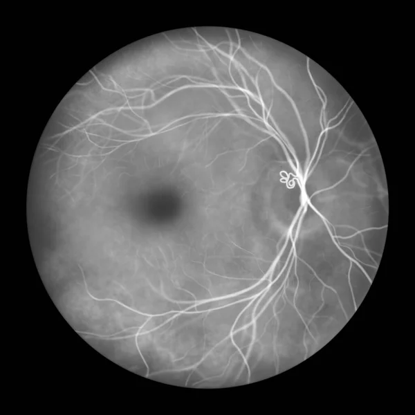
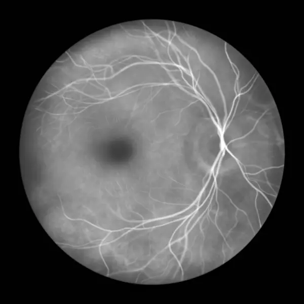
Stock image A prepapillary vascular loop on the retina, as observed during ophthalmoscopy in fluorescein angiogram, an illustration showcasing the looping blood vessels around the optic disc.
Published:
Sep.26, 2023 14:25:47
Author:
katerynakon
Views:
2
Downloads:
1
File type:
image
/
jpg
File size:
2.77 MB
Orginal size:
5000
x
5000
px
Available sizes:
S
(500 x 500)
M
(1000 x 1000)
L
(2000 x 2000)
XL
(5000 x 5000)
EL0
(5000 x 5000)
Level:
silver
Download Image
Image topics:
Illustration
Health
Blood
Medical
Eye
Drawing
Variation
Loops
Optic
Disc
Examination
Anatomy
Loop
Ophthalmic
Ocular
Ophthalmology
Vessels
Conditions
Diseases
Abnormal
Diagnostics
Congenital
Vascular
Pathology
Variations
Retina
Nerve
Ophthalmoscopy
Retinal
Disorders
Capillaries
Angiogram
Ophthalmoscope
Fundus
Hand Drawn
Eye Examination
Vasculature
Angiography
Medical Illustration
Optic Disc
Anomalies
Retinoscopy
Abnormalities
Fundoscopy
Fluorescein
Prepapillary Vascular Loop
Prepapillary
Retinal Vasculature
Vascular Anomalies
Retinal Anatomy
Similar stock images
Retinal Arteriovenous Malformation, Rare Congenital Retinal Vascular Anomalies. An Illustration Shows Artery-vein Communication Without Intervening Capillaries In Fluorescein Angiography.
5000 × 5000
A Prepapillary Vascular Loop On The Retina, As Observed During Ophthalmoscopy In Fluorescein Angiogram, An Illustration Showcasing The Looping Blood Vessels Around The Optic Disc.
5000 × 5000
Medical Illustration Of A Submacular Hemorrhage Observed In A Fluorescein Angiogram, Showcasing A Dark, Irregularly Shaped Hemorrhage Within The Macular Region Of The Retina.
5000 × 5000
Normal Retina, Illustration Of An Ophthalmoscope Image In Fluorescein Angiography.
5000 × 5000
Non-proliferative Diabetic Retinopathy, Illustration Showing Venous Beading, Ophthalmoscope View, Fluorescein Angiography
5000 × 5000
Normal Eye Retina, Ophthalmoscope View, Scientific Illustration Showing Optic Disk, Blood Vessels, Macula And Fovea, Fluorescein Angiography
5000 × 5000
Diabetic Macular Edema (DME), Illustration Shows Cystoid Macula Edema And Optic Disk Edema, Abnormal Finding On Fundoscopic Examination Of The Eye Retina In Diabetes Mellitus, Fluorescein Angiography
5000 × 5000
Normal Eye Retina, Scientific Illustration Showing Optic Disk, Blood Vessels, Macula And Fovea, Fluorescein Angiography, Digital Illustration
5000 × 5000