
Stock image Protein Interaction
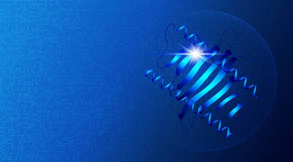
Proteomics And Functional Genomics - The Large-scale Study Of Proteins In Living Organisms - A Protein Isolated On Blue Tech Background - Conceptual Illustration With Copy Space
Image, 10.29MB, 9584 × 5298 jpg

Proteomics And Protein Folding Prediction Through Computational Means - The Study Of The Function And Structure Of Proteins Within Living Organisms - Conceptual Illustration
Image, 10.62MB, 6500 × 3656 jpg
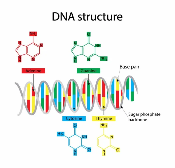
Illustration Of Biology, DNA Structure Consists Of Two Strands Of Nucleotides That Are Held Together By Hydrogen Bonds, Four Nitrogenous Bases: Adenine , Thymine, Guanine, Cytosine
Vector, 5.49MB, 4186 × 4024 eps
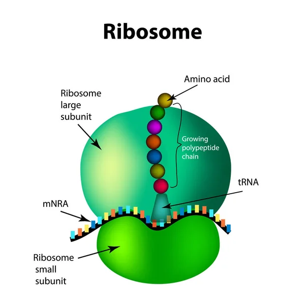
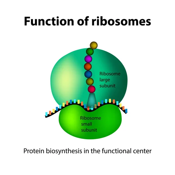
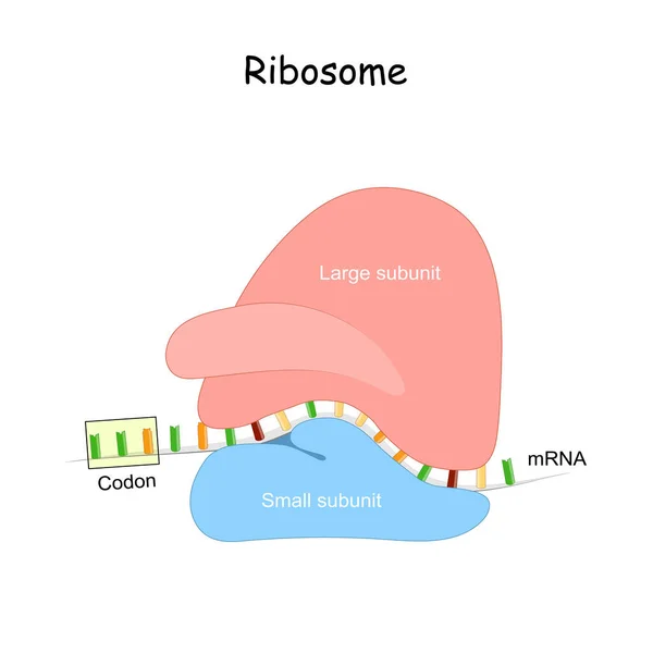
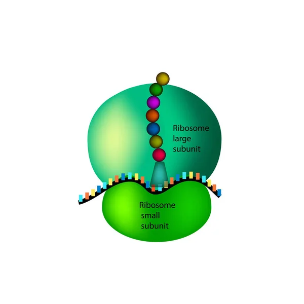
The Structure Of The Ribosome. Infographics. Vector Illustration On Isolated Background
Vector, 2.01MB, 5000 × 5000 eps
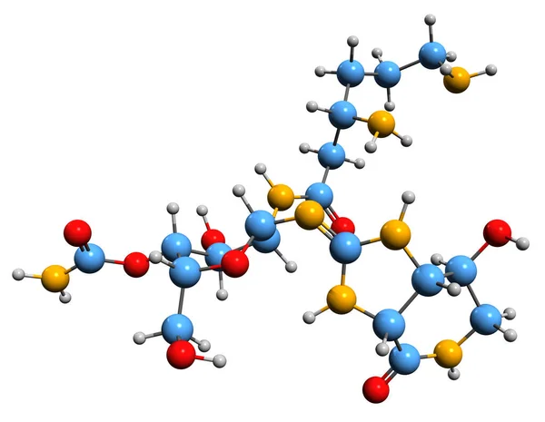
3D Image Of Nourseothricin Skeletal Formula - Molecular Chemical Structure Of Aminoglycoside Antibiotic Isolated On White Background
Image, 5.03MB, 7855 × 6096 jpg

The Structure Of The Ribosome. Functions. Infographics. Vector Illustration On Isolated Background
Vector, 1.89MB, 5000 × 5000 eps

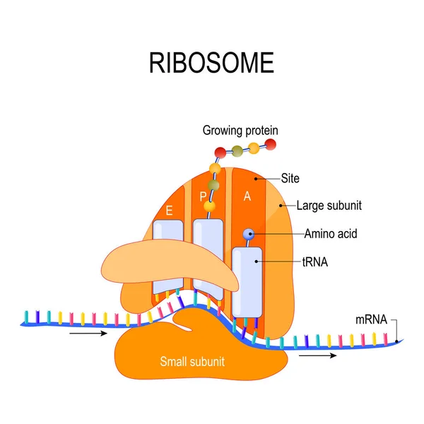
Anatomy Of A Ribosome. The Interaction Of A Ribosome With MRNA. Process Of Initiation Of Translation (biological Protein Synthesis). Vector Diagram For Your Design, Educational, Medical, Biological And Science Use
Vector, 0.77MB, 4452 × 4452 eps
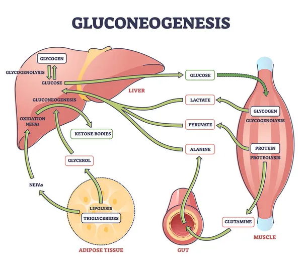
Gluconeogenesis GNG Metabolic Pathway For Glucose Generation Outline Diagram
Vector, 6.31MB, 4500 × 3938 eps

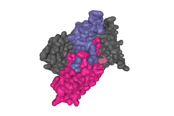
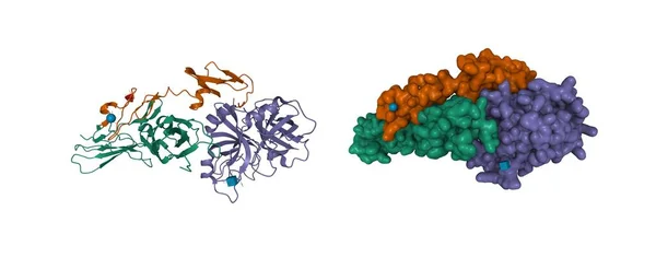
Structure Of Follicle-stimulating Hormone (color) In Complex With The Entire Ectodomain Of Its Receptor (grey), 3D Gaussian Surface Model, White Background
Image, 0.75MB, 6000 × 4000 jpg


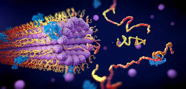
Ribosome And MRNA. Anatomy Of Macromolecular Machines, That Protein Synthesis. MRNA Translation. Small And Large Subunit. MRNA Vaccine. Explained Biology Basics.
Vector, 0.7MB, 4444 × 4444 eps


Integrin Alpha2 I Domain (green) In Complex With Collagen, 3D Cartoon Model, White Background
Image, 2.52MB, 6000 × 4000 jpg


Structure Of Tissue Factor (green) -factor VIIa (brown And Violet) Complex, 3D Cartoon And Gaussian Surface Models, Chain Id Color Scheme, Based On PDB 1j9c, White Background
Image, 3.57MB, 10000 × 4100 jpg


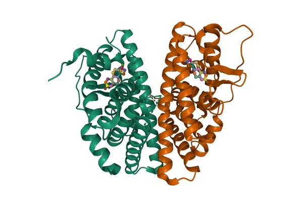
Estrogen Receptor Beta Dimer In Complex With Estradiol, 3D Cartoon Model, Chain Id Color Scheme, Based On PDB 5toa, White Background
Image, 2.91MB, 6000 × 4000 jpg


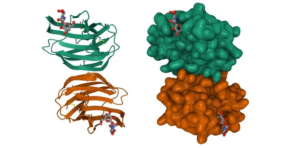
Crystal Structure Of Human Galectin-1 In Complex With Type 1 N-acetyllactosamine. 3D Cartoon And Gaussian Surface Model, Chain Id Color Scheme, PDB 4xbl, White Background
Image, 3.8MB, 8000 × 4000 jpg
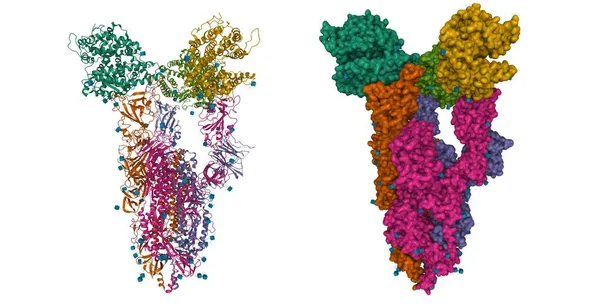
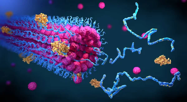
Structure Of The SARS-CoV-2 Spike Glycoprotein, Surface Model, White Background, 3D Illustration Isolated
Image, 0.81MB, 5000 × 3380 jpg

Eggs Painted With Emotions, Psychology, Feelings, Communication And Perception White Background
Image, 1.56MB, 5000 × 3312 jpg

Crystal Structure Of SARS-CoV-2 Nsp16 (green)-nsp10 (brown)-ligand (ball-and-stick) Complex, 3D Cartoon Model, White Background
Image, 2.07MB, 4096 × 4096 jpg
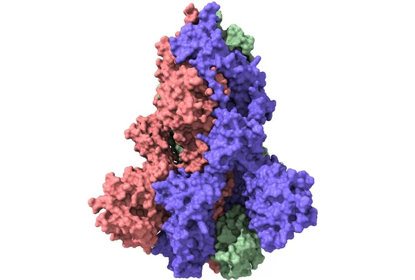
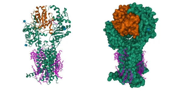
Structure Of Triple ACE2-bound SARS-CoV-2 Trimer Spike, 3D Cartoon And Gaussian Surface Models, White Background
Image, 6.77MB, 8000 × 4100 jpg


Structure Of The SARS-CoV-2 Spike Glycoprotein, Surface Model, Black Background, 3D Illustration Isolated
Image, 0.93MB, 5000 × 3380 jpg
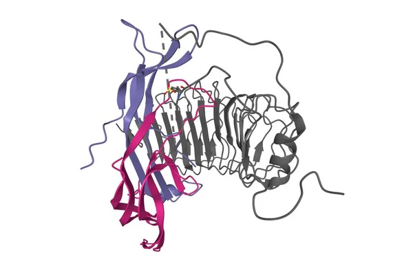
Structure Of Follicle-stimulating Hormone (color) In Complex With The Entire Ectodomain Of Its Receptor (grey), 3D Ribbon Model, White Background
Image, 1.39MB, 6000 × 4000 jpg


Structure Of Murine Dispatched (green) In Complex With Native Sonic Hedgehog (brown). Cell Membrane Cholesterol Is Shown In Pink. 3D Cartoon And Gaussian Surface Models, PDB 7rpk, White Background
Image, 5.23MB, 8000 × 4000 jpg
Protein Enzymes Fold Into Their Structure To Fulfill Their Function - 3d Illustration
Image, 13.4MB, 7300 × 4000 jpg
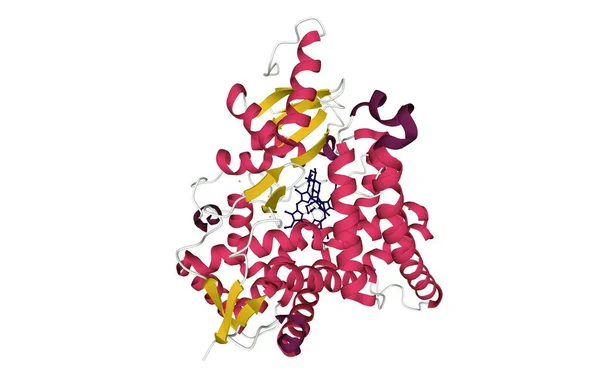
Human Placental Aromatase Cytochrome P450 (CYP19A1) Complexed With Testosterone, 3D Cartoon Model With Colored Elements Of The Secondary Structure, White Background
Image, 2.33MB, 6258 × 3993 jpg

The Structure Of The Ribosome. Infographics. Vector Illustration On Isolated Background
Vector, 1.85MB, 5000 × 5000 eps
Protein Enzymes Fold Into Their Structure To Fulfill Their Function - 3d Illustration
Image, 10.02MB, 7300 × 3500 jpg
Crystal Structure Of A Complex Formed Between Group II Phospholipase A2 (green) And Aspirin, 3D Model, Black Background
Image, 4.54MB, 6077 × 3993 jpg
Page 1 >> Next