
Stock image Short Pr Interval
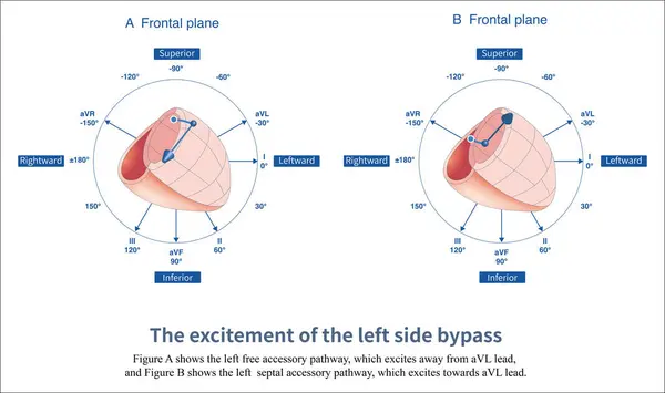
When The Left Free Wall And Septal Accessory Pathway Are Excited, Preexcitation Waves With Different Polarities Are Generated In Leads And AVL.
Image, 6.98MB, 14851 × 8810 jpg

Some Accessory Pathways Directly Connect Atrium With The Lower Part Of Atrioventricular Node And His Bundle, Bypassing The Slow Conduction Area Of Atrioventricular Node.
Image, 10.59MB, 7000 × 13649 jpg

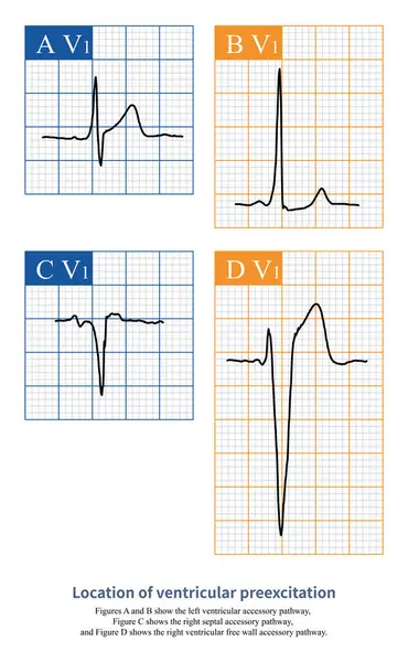
On The Electrocardiogram, Observing The Morphology Of QRS Waves In Lead V1 Can Distinguish Whether Ventricular Pre Excitation Is Located In The Left Ventricle Or The Right Ventricle.
Image, 4.78MB, 10000 × 11226 jpg

When The Ventricular Preexcitation Wave Leaves The Baseline And Then Falls Back To The Baseline, It Is Interpreted As An Isoelectric Line Preexcitation Wave.
Image, 9.3MB, 10000 × 11275 jpg

When The Left Anterior Wall And Posterior Wall Accessory Pathway Are Excited, Preexcitation Waves With Different Polarities Are Generated In The Inferior Wall Leads Of , And AVF.
Image, 3.66MB, 10000 × 5932 jpg

Ventricular Preexcitation Is The Pre Excitation Of A Portion Of The Ventricular Muscle By The Accessory Pathway, Forming A Rough And Dull, And Fuzzy Wave That Can Be Positive, Negative, Or Biphasic.
Image, 7.55MB, 10000 × 11891 jpg

Ventricular Preexcitation Is A Fusion Wave Formed By The Accessory Pathway And Normal Atrioventricular Conduction System Exciting A Part Of Ventricle Respectively.
Image, 4.25MB, 10000 × 10000 jpg

When Ventricular Preexcitation Components Occupy Different Proportions Of QRS Waves, The Measured PJ Intervals Are Different.
Image, 4.21MB, 10000 × 4226 jpg

On The Electrocardiogram, Observing The Morphology Of QRS Waves In Lead V1 Can Distinguish Whether Ventricular Pre Excitation Is Located In The Left Ventricle Or The Right Ventricle.
Image, 18.67MB, 10000 × 14900 jpg

When A Short PR Interval Is Found On The Electrocardiogram, Careful Observation Of The Beginning Of The QRS Wave Can Reveal Whether It Is Ventricular Preexcitation.
Image, 8.71MB, 10000 × 7702 jpg
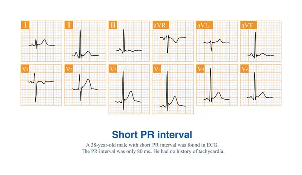
The PR Interval Of An Adult ECG Is Less Than 120 Ms, Which Is Called A Short PR Interval.
Image, 10.02MB, 10000 × 5814 jpg

The Polarity Of Ventricular Preexcitation Waves Can Be Positive, As Shown In Figures A And B, Or Negative, As Shown In Figures C And D.
Image, 2.82MB, 10000 × 11593 jpg

When There Is A Left Ventricular Free Wall Bypass, The Polarity Of The Ventricular Preexcitation Is Positive In Lead V1 And Negative In Lead AVL On The Electrocardiogram.
Image, 7.36MB, 10000 × 6759 jpg

On The Electrocardiogram, Observing The Morphology Of QRS Waves In Lead V1 Can Distinguish Whether Ventricular Pre Excitation Is Located In The Left Ventricle Or The Right Ventricle.
Image, 2.99MB, 10000 × 6863 jpg

At The Beginning Of Heart Development, The Atria And Ventricles Form A Whole, Known As The Primitive Heart Tube.
Image, 4.96MB, 10000 × 10005 jpg
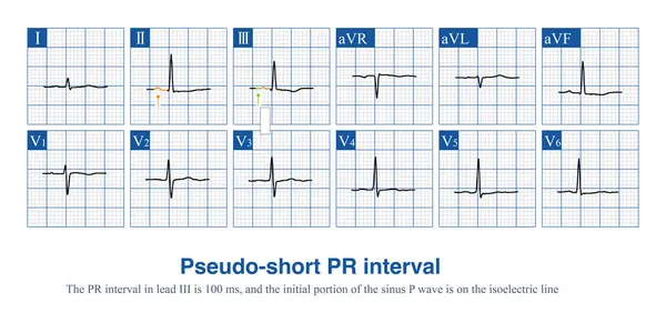
Sometimes, The Beginning Of Sinus P Wave In Some ECG Leads Is Located On The Isoelectric Line. If Only The Visualization Part Is Measured, It Will Produce A Pseudo Short PR Interval.
Image, 9.68MB, 10000 × 4859 jpg
On The Electrocardiogram, Observing The Morphology Of QRS Waves In Lead V1 Can Distinguish Whether Ventricular Pre Excitation Is Located In The Left Ventricle Or The Right Ventricle.
Image, 7.46MB, 8000 × 12972 jpg

When Individuals With Short PR Interval ECG Develop Various Types Of Supraventricular Tachycardia Clinically, It Is Called Short PR Syndrome.
Image, 8.01MB, 11811 × 5069 jpg
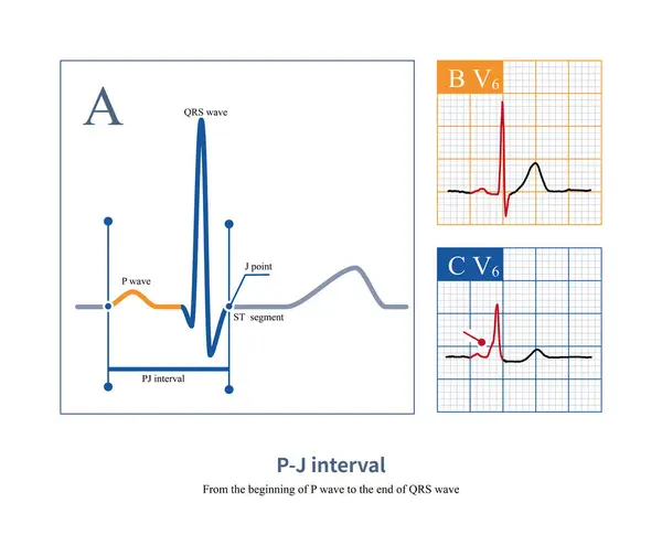
On The Electrocardiogram, The PJ Interval Is Used To Distinguish Between Ventricular Heartbeats And Ventricular Pre Excitation. The Normal Value Of PJ Interval Is Less Than 270ms.
Image, 8.38MB, 10000 × 8409 jpg
Page 1 >> Next