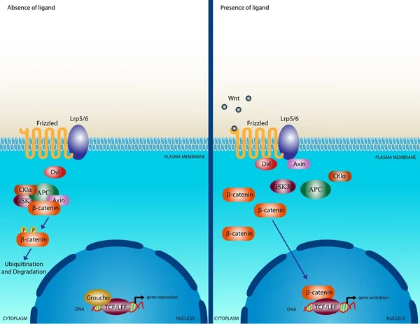
Stock image Some accessory pathways directly connect atrium with the lower part of atrioventricular node and His bundle, bypassing the slow conduction area of atrioventricular node.

Published: Jun.29, 2024 09:10:25
Author: asia11m
Views: 0
Downloads: 0
File type: image / jpg
File size: 10.59 MB
Orginal size: 7000 x 13649 px
Available sizes:
Level: beginner