
Stock image Signal Cascade
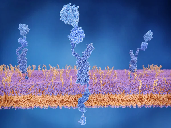
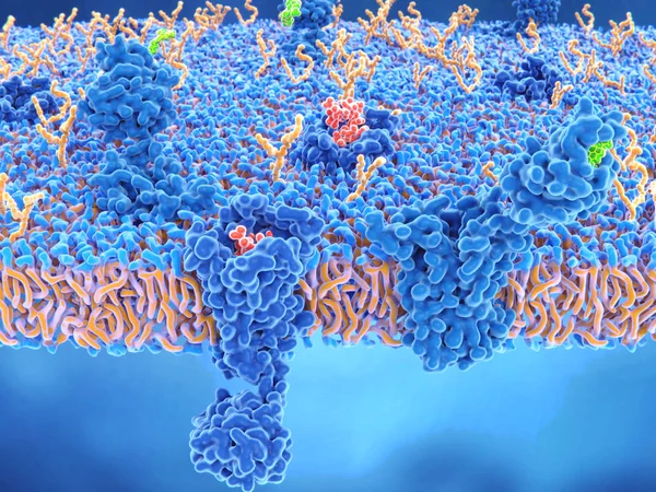
The Amyloid Precursor Protein (APP) Is A Complex Protein With Many Functions. It Is Found On The Surface Of Cells Throughout The Body. The Intact Protein Binds To Many Stuctural Proteins Outside Cells, Such As Heparin And Laminin And Sends Signals Th
Image, 6.49MB, 8000 × 6000 jpg
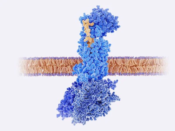
The Calcitonin Gene Related Peptide (yellow) Binds To Its Receptor (blue) On Neurons And Smooth Muscle Cells Of Cerebral Blood Vessels, Activating A Signal Cascade Through G-proteins (dark Blue) In This Cells That Leads To A Dilatation Of The Blood
Image, 4.43MB, 8000 × 6000 jpg
Rhodopsin Is A Light Sensitive G-protein Coupled Receptor With Retinal As Cofactor. That Stimulates The G-protein Transducin, Resulting In The Liberation Of Its Subunit. This GTP-bound Subunit In Turn Activates CGMP Phosphodiesterase.
Image, 8.93MB, 8000 × 6000 jpg

Interactions Of MHC-II With The T-cell Receptor And CD4 And B7-1 With CD-28 Activates T-cells While The Interactions Of P7-1 With CTLA-4 And PD-L1 With PD-1 Deactivates T-cells.
Image, 10.7MB, 8000 × 6000 jpg

Dopamine And The Dopamine Receptor D1. The Dopamine Binding Side Is Insinuated. Source: PDB Entry 7ljd
Image, 6.96MB, 8000 × 6000 jpg

The Calcitonin Gene Related Peptide (yellow) Binds To Its Receptor (blue) On Neurons And Smooth Muscle Cells Of Cerebral Blood Vessels, Activating A Signal Cascade Through G-proteins (dark Blue) In This Cells That Leads To A Dilatation Of The Blood
Image, 7.55MB, 8000 × 6000 jpg
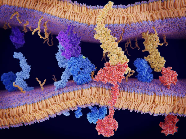
PD-1 (red) Extends From The Surface Of A T-cell And Interacts With The Ligand Protein PD-L1 (yellow) From A Antigen Presenting Cell. Although The T-cell Has Been Activated Through The Interaction Of A T-cell Receptor (blue) And A MHC Protein (viole
Image, 18.32MB, 8000 × 6000 jpg

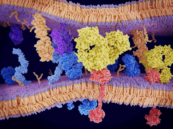
Activation Of The Immune Response To An Antigene (green) Through The Complex Between A T-cell Receptor (dark Blue), An MHC II-antigen (violet) And A CD4 Protein (light Blue). 3d Rendering. Illustration
Image, 6.36MB, 8000 × 6000 jpg

The Opioid Receptors Display A Role In Modulating Pain Perception; Opioid Agonists Are Therefore Potent Analgesics. They Appear Mainly In The Brain And Spinal Cord. The Endogenous Opioids Are Enkephalin, Dynorphin, Endorphin And Nociceptin.
Image, 7.96MB, 8000 × 6000 jpg
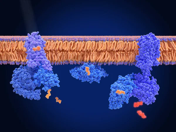
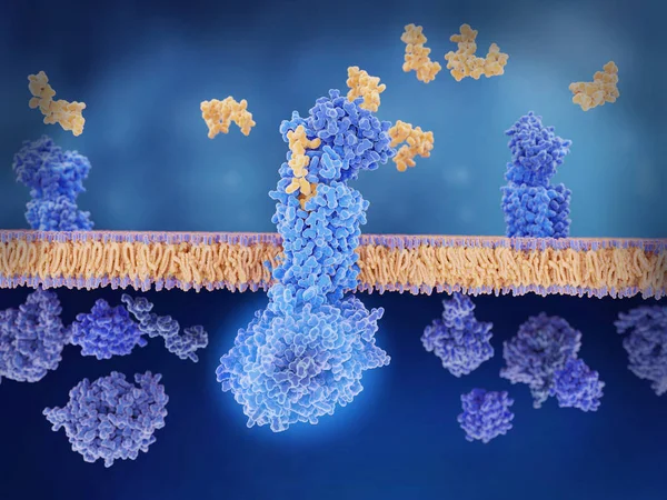
Cancer Cells Express PD-L1 (orange) Proteins On Their Surface To Trick The Immune System. The Interaction Of PD-L1 With PD-1 Of T-cells Leads To A Down-regulation Of T-cells. The Antibody (yellow) Blocks This Interaction.
Image, 18.3MB, 8000 × 6000 jpg
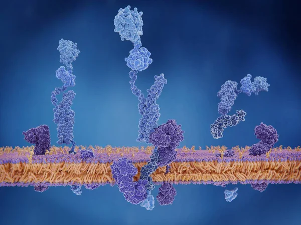
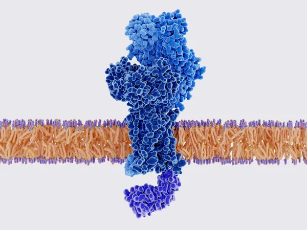
The Amyloid Precursor Protein. When Cleaved, The Membrane Domain Is Involved In The Alzheimer Disease Building Amyloid Plaques. 3d Rendering. Illustration
Image, 3.29MB, 8000 × 6000 jpg

Activation Of The GABA B Receptor By Baclofen. GABA B Receptors Are G Protein-coupled Receptors. Binding Of An Agonist (baclofen, Red) Leads To A G-protein Coupled C-AMP Signaling Pathway. Source: PDB Entries 7eb2, 6r3q,.
Image, 9.97MB, 8000 × 6000 jpg
The Calcitonin Gene Related Peptide (yellow) Binds To Its Receptor (blue) On Neurons And Smooth Muscle Cells Of Cerebral Blood Vessels, Activating A Signal Cascade Through G-proteins (dark Blue) In This Cells That Leads To A Dilatation Of The Blood
Image, 6.01MB, 8000 × 6000 jpg
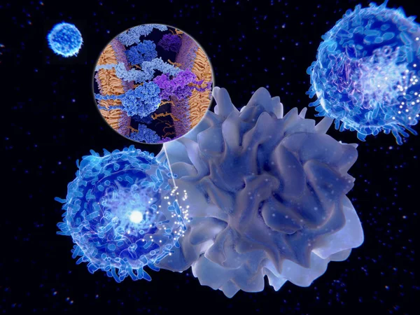
Dendritic Cells Present Antigens (green) To Lymphocytes Through Their Membran Bound MHC-molecules (violet). CD4 Molecules (light Blue) Bind To Other Portions Of The MHC, Strengthening The Interaction.
Image, 10.24MB, 8000 × 6000 jpg
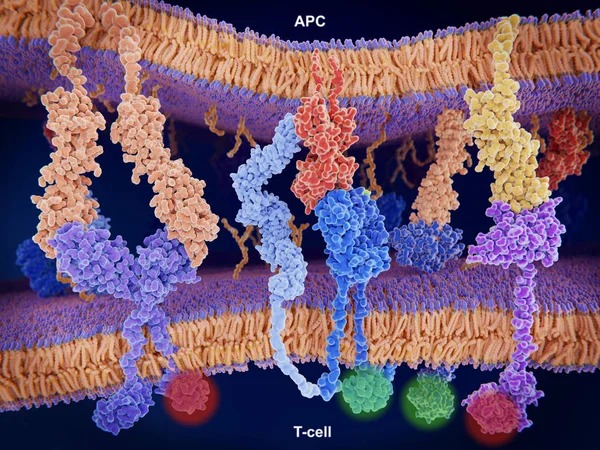
Interaction Of MHC-II (red) With The T-cell Receptor (blue) And CD4 (light Blue) And B7-1 (orange) With CD-28 (dark Blue) Activates T-cells While The Interaction Of P7-1 With CTLA-4 (violett) And PD-L1 (yellow) With PD-1 Deactivates T-cells
Image, 10.65MB, 8000 × 6000 jpg
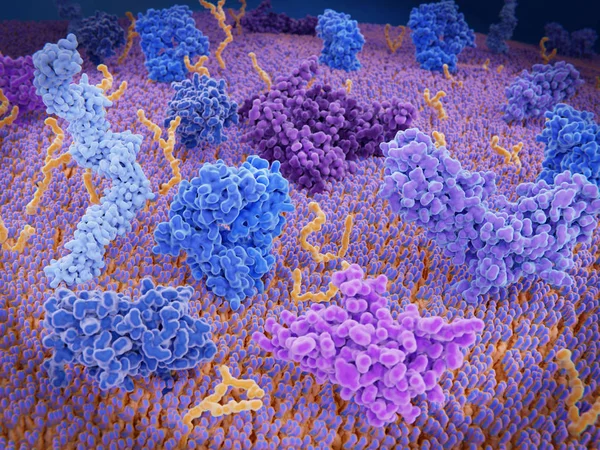
The T-cell Receptor Activates The Immune Response To Antigens In T-lymphocytes. T-cell Receptors (dark Blue), CD4 Molecules (light Blue), Glycolipids (orange). 3d Rendering. Illustration
Image, 3.11MB, 8000 × 6000 jpg
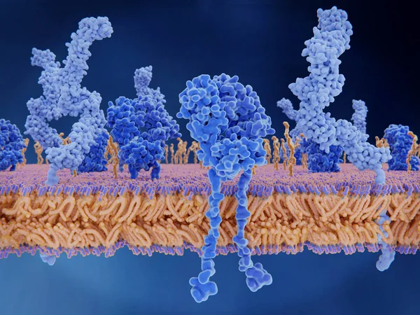
Immunologically Active Proteins On A T-cell. TCR (blue), CD-4 (light Blue), CD-28 (dark Blue), PD-1 (magenta), CTLA-4 (violet), Ca-channel (dark Violet). The T-cell Receptor, CD-4 And CD-28 Activate T-cells, While PD-1 And CTLA-4 Inhibit The Activat
Image, 10.2MB, 8000 × 6000 jpg
The Opioid And Cannabinoid Receptors Are Involved In Pain-sensation, Mood, Appetite And Memory. Agonists Are Potent Analgesics: Endorphin (red) And Tetrahidrocannabinol (green).
Image, 8.93MB, 8000 × 6000 jpg

Cyclic Adenosine Monophosphat (cAMP, Red) Is A Second Messenger Used For Signal Transduction Through The Activation Of Various Protein Kinases (blue). The One In The Foreground Is Protein Kinase A. Source: PDB Entry 3tnp.
Image, 7MB, 8000 × 6000 jpg
Control UI Pixel Perfect Well-crafted Vector Thin Line And Solid Icons
Vector, 0.59MB, 4544 × 2064 eps
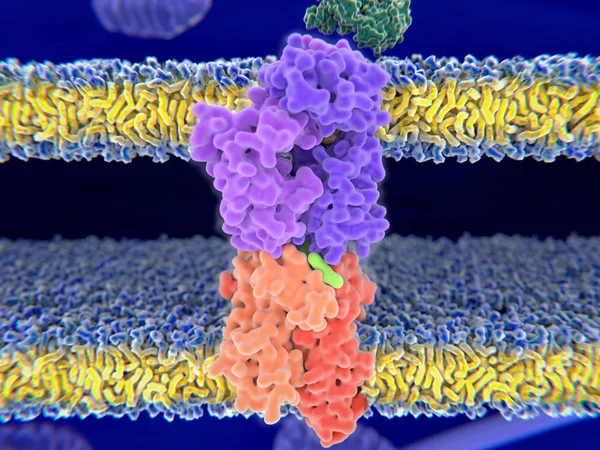
T-cell Receptor In Complex With The MHC Class II-peptide Complex. The Antigen (light Green) Is A Peptide From A Tumor Cell, Bacteria Or Virus. Complex Embedded In The Membranes. 3D-Rendering. Illustration
Image, 7.59MB, 8000 × 6000 jpg
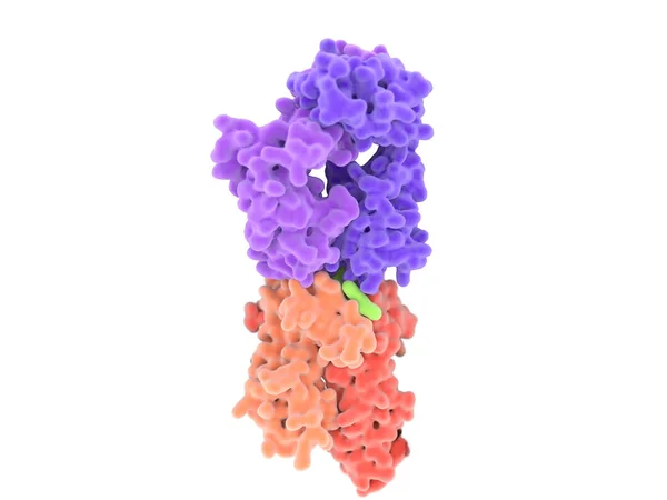
T-cell Receptor In Complex With The MHC Class II-peptide Complex. The Antigen (light Green) Is A Peptide From A Tumor Cell, Bacteria Or Virus. Different Stages Of The Interaction. 3D-Rendering. Illustration
Image, 2.17MB, 8000 × 6000 jpg

Nausea Vector Illustration. Labeled Medical Vomiting Explanation Scheme.
Vector, 8.95MB, 4500 × 3830 eps


Unread Messages Organized As A Cascade Of Pages Popping Out Of The Laptop, Overwhelming Work
Vector, 0.28MB, 10000 × 7083 eps
Page 1 >> Next