
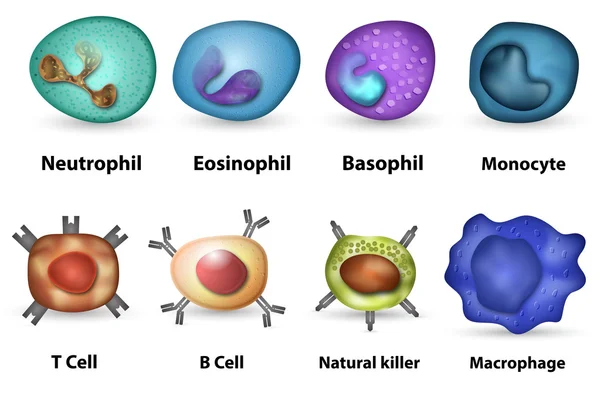
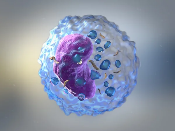
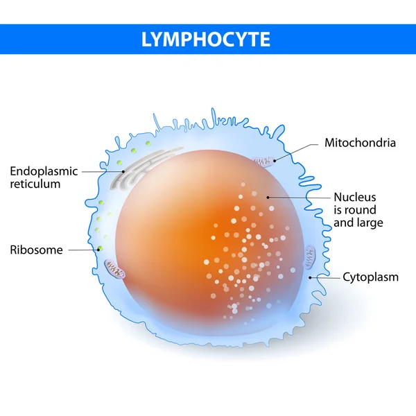
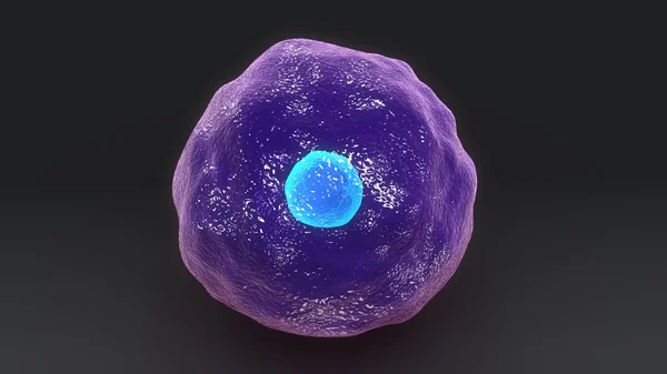
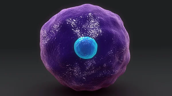
Stock image T Lymphocyte
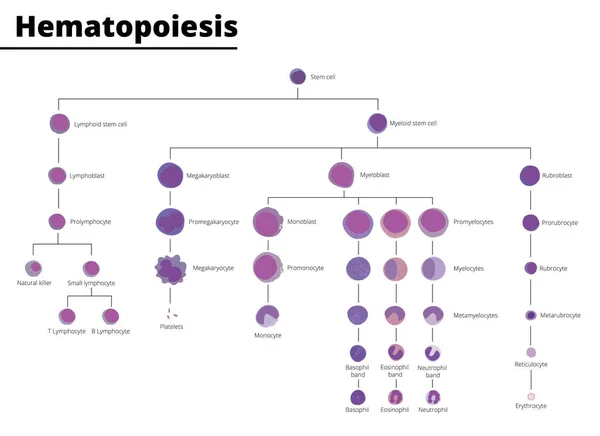
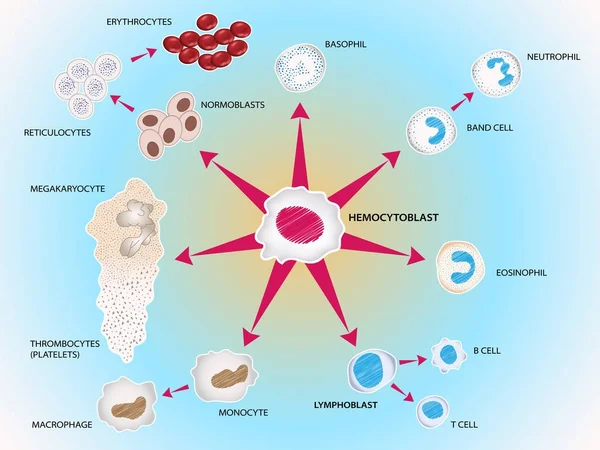
Hematopoiesis Differentiation Of Blood Cell Types Infographic Stem Cell Derived Blood Cells And Immune Cells. Vector Illustration. Didatic Illustration.
Vector, 1.2MB, 5500 × 4000 ai

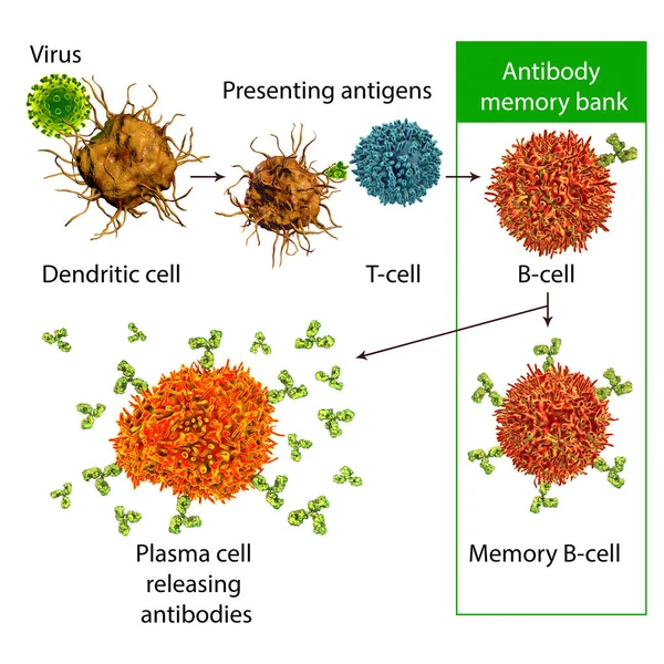
B-cell Leukocyte Activation By Antigen. From Antigen Binding To B Cell Receptor, And Chemical Signal Of T-cell Helper To Becomes Plasma Cell And Antibodies Releases. White Blood Cell. Vector Illustration
Vector, 5.19MB, 11801 × 8257 eps
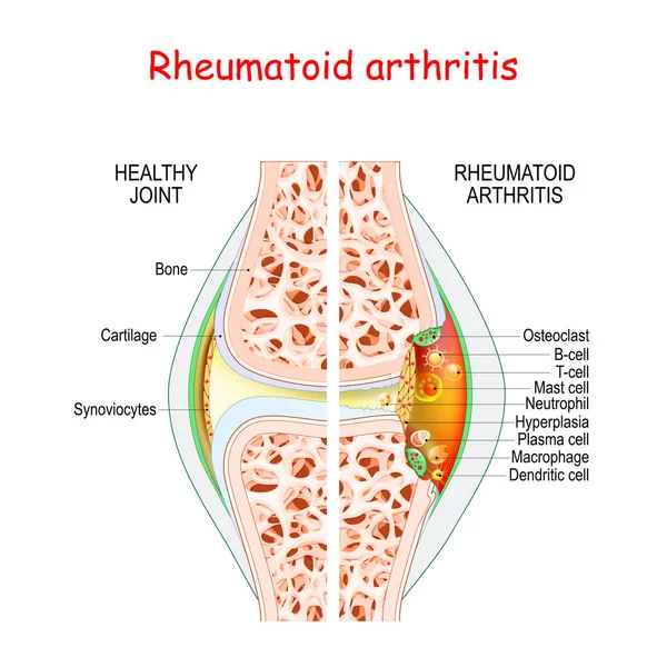
Rheumatoid Arthritis. Healthy And Damage Joint. Close-up Of Bone, Cartilage, And Cells In A Joint Capsule: Synoviocytes, Osteoclast, Neutrophil, T Lymphocyte, B-cell, Macrophage, Mast, Plasma, And Dendritic Cell,
Vector, 12.31MB, 4444 × 4444 eps
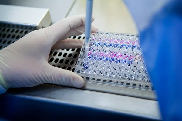
Laboratory Developing Therapeutic Vaccines For The Treatment Of Lung Cancer By Stimulating The Immune System.
Image, 16.14MB, 5616 × 3744 jpg
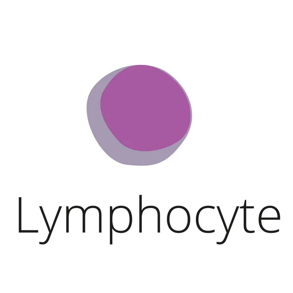
Lymphocyte B And T-cell. Cells Of Immune System (immune Response). Vector Illustration On White Background. Didatic Illustration.
Vector, 0.49MB, 5000 × 5000 ai


The Life Cycle Of HIV. Infographics. World AIDS Day. Vector Illustration
Vector, 10.19MB, 5000 × 5000 eps
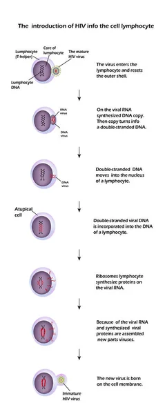
The Life Cycle Of HIV. Infographics. World AIDS Day. Vector Illustration
Vector, 9.28MB, 3276 × 8190 eps
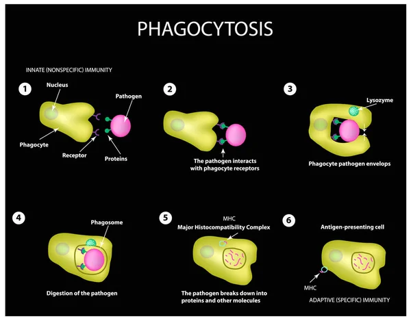
Innate Immunity. Adaptive Specific . Phagocytosis. Infographics. Vector
Vector, 3.99MB, 5000 × 3911 eps
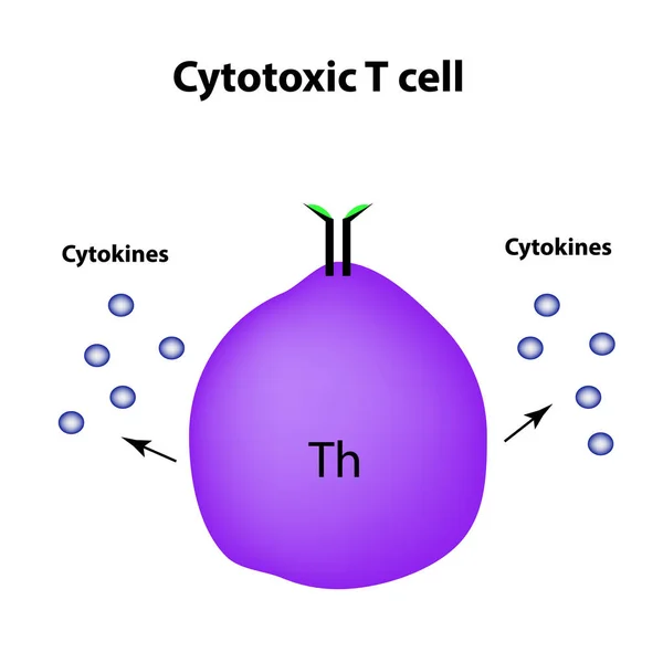
Cytotoxic Cells. Cytokines. Cell Immunity. Infographics. Vector Illustration
Vector, 1.39MB, 5000 × 5000 eps
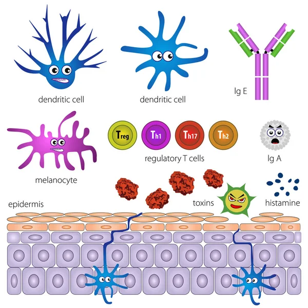
The Cells Of The Protective Layer Of The Skin. Protection Against Inflammation
Vector, 1.63MB, 5000 × 5000 eps
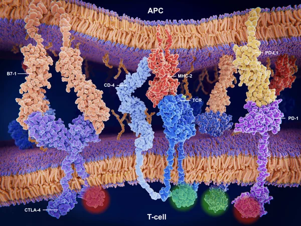
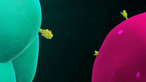
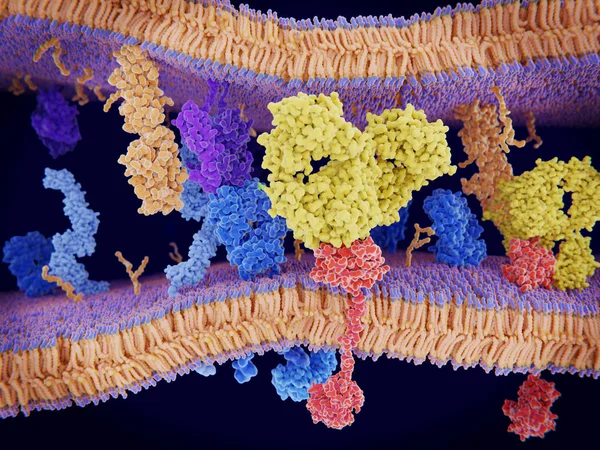
Interactions Of MHC-II With The T-cell Receptor And CD4 And B7-1 With CD-28 Activates T-cells While The Interactions Of P7-1 With CTLA-4 And PD-L1 With PD-1 Deactivates T-cells.
Image, 10.7MB, 8000 × 6000 jpg


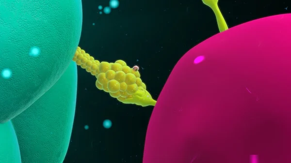
Activation Of The Immune Response To An Antigene (green) Through The Complex Between A T-cell Receptor (dark Blue), An MHC II-antigen (violet) And A CD4 Protein (light Blue). 3d Rendering. Illustration
Image, 6.36MB, 8000 × 6000 jpg
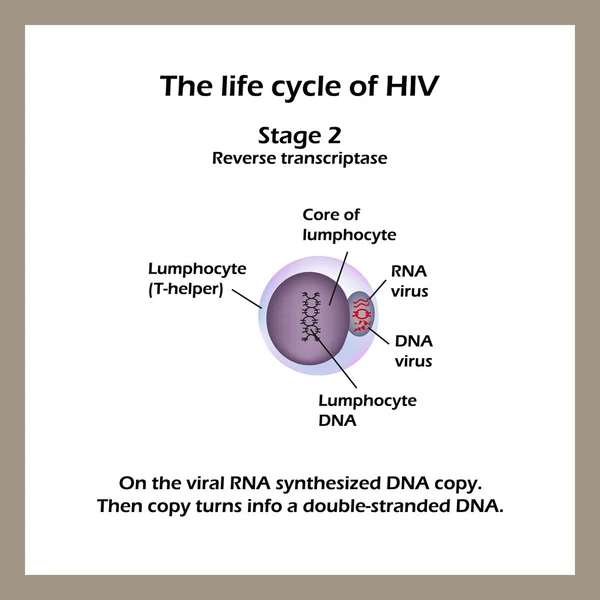
The Life Cycle Of HIV. Stage 2 -The Viral RNA Synthesized DNA Copy. World AIDS Day. Vector Illustration
Vector, 3.88MB, 5000 × 5000 eps
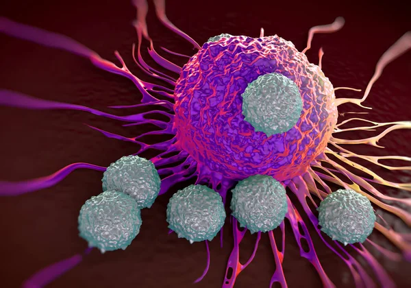
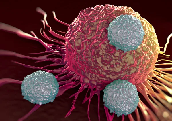
Cancer Cells Express PD-L1 (orange) Proteins On Their Surface To Trick The Immune System. The Interaction Of PD-L1 With PD-1 Of T-cells Leads To A Down-regulation Of T-cells. The Antibody (yellow) Blocks This Interaction.
Image, 18.3MB, 8000 × 6000 jpg
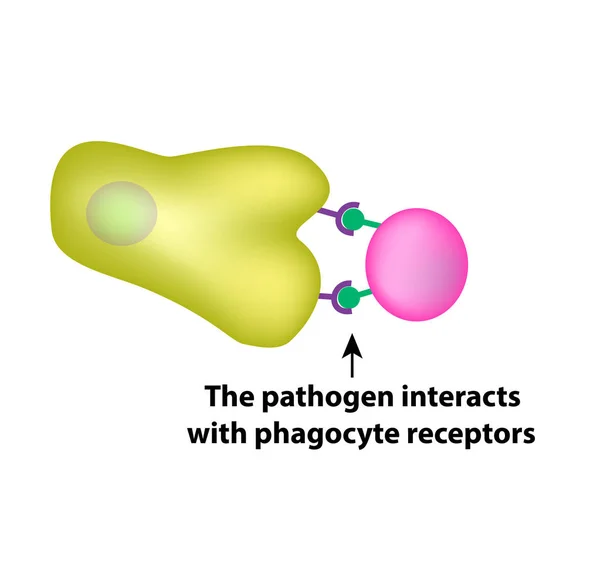
Innate Immunity. Adaptive Specific . Phagocytosis. Infographics. Vector Illustration
Vector, 1.45MB, 5000 × 4734 eps
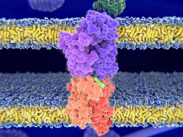
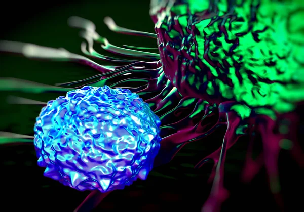
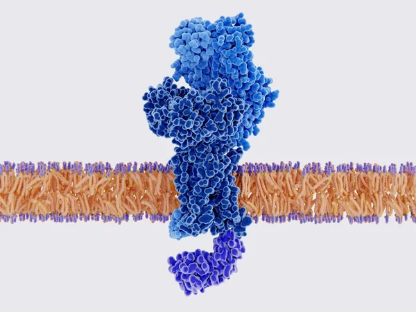
T-cell Receptor In Complex With The MHC Class II-peptide Complex. The Antigen (light Green) Is A Peptide From A Tumor Cell, Bacteria Or Virus. Complex Embedded In The Membranes. 3D-Rendering. Illustration
Image, 7.59MB, 8000 × 6000 jpg
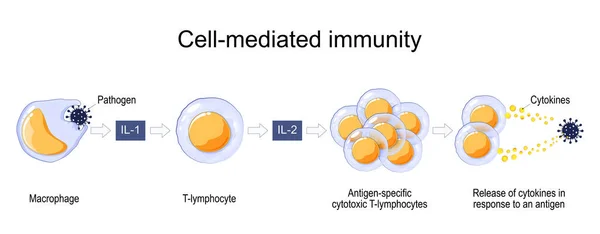
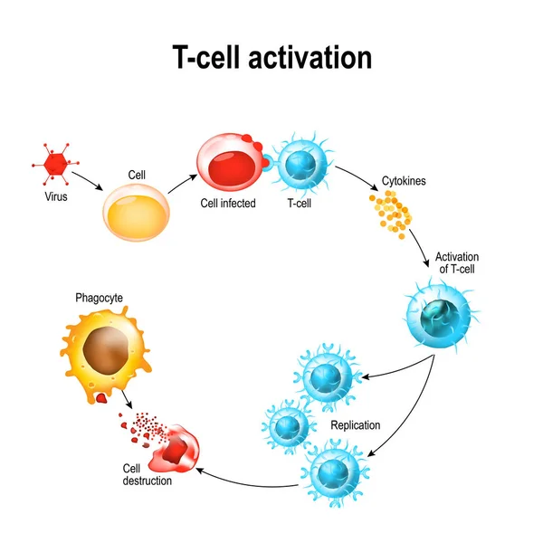
Immune Response. Cell-mediated Immunity. Activation Of Phagocytes, Antigen-specific Cytotoxic T-lymphocytes, And The Release Of Cytokines In Response To An Antigen. Vector Poster For Educatio
Vector, 11.15MB, 7000 × 2734 eps
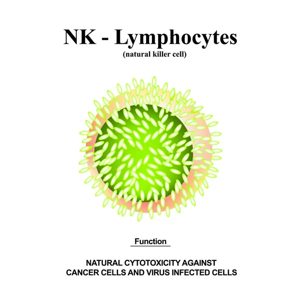
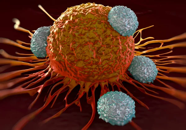
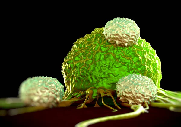
NK Lymphocytes Structure. The Functions Of NK Lymphocytes. Immunity Helper Cells. Infographics. Vector Illustration On Isolated Background.
Vector, 8.18MB, 5000 × 5000 eps
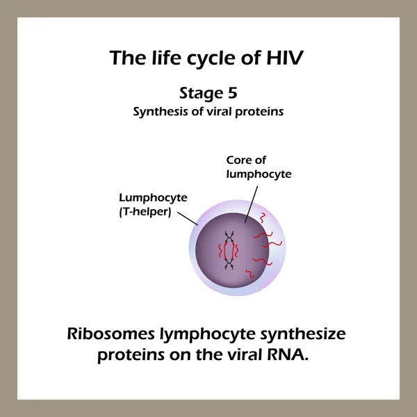
The Life Cycle Of HIV. Stage 5 - Ribosomes Lymphocyte Cells Synthesize Proteins In The Virus RNA. World AIDS Day.
Vector, 4.11MB, 5000 × 5000 eps

T-cell Receptor In Complex With The MHC Class II-peptide Complex. The Antigen (light Green) Is A Peptide From A Tumor Cell, Bacteria Or Virus. Different Stages Of The Interaction. 3D-Rendering. Illustration
Image, 7.31MB, 8000 × 6000 jpg

The Life Cycle Of HIV. Stage 1 - The Virus Enters The Lymphocyte. Vector Illustration
Vector, 4.32MB, 5000 × 5000 eps
Page 1 >> Next