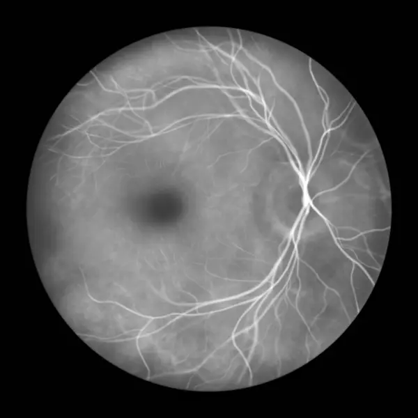
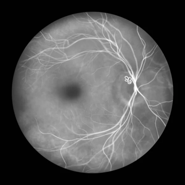
Stock image Medical illustration of a submacular hemorrhage observed in a fluorescein angiogram, showcasing a dark, irregularly shaped hemorrhage within the macular region of the retina.

Published: Sep.26, 2023 14:25:47
Author: katerynakon
Views: 4
Downloads: 0
File type: image / jpg
File size: 2.77 MB
Orginal size: 5000 x 5000 px
Available sizes:
Level: silver