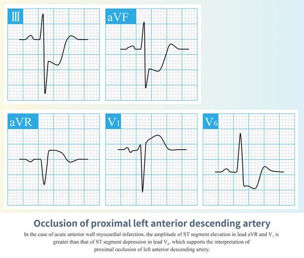
Stock image After the first diagonal branch and before the first septal branch of the LAD were occluded, the infarction affected the anteroseptum, and the high lateral wall was not affected.

Published: Mar.09, 2023 15:09:46
Author: asia11m
Views: 47
Downloads: 0
File type: image / jpg
File size: 17.3 MB
Orginal size: 10000 x 8459 px
Available sizes:
Level: beginner