Home
Images
Vectors
Videos
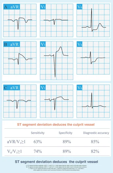
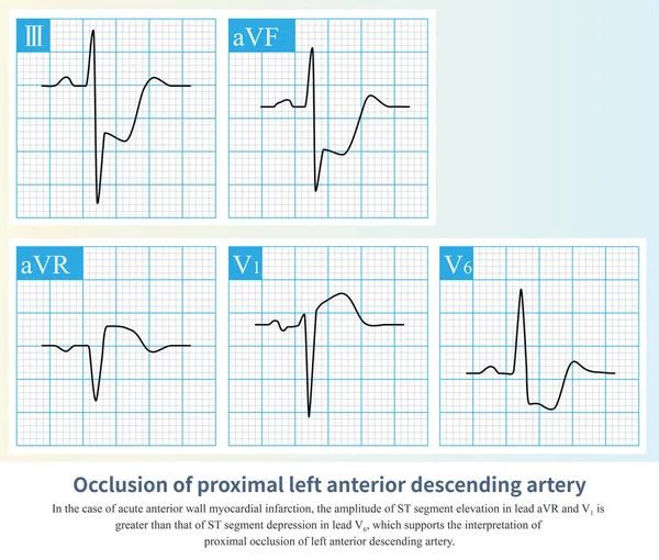
Stock image In the case of acute anterior myocardial infarction, the amplitude of ST segment offset can be used to deduce that the occlusive site is located in the proximal segment of the LAD.
Published:
Mar.06, 2023 10:29:20
Author:
asia11m
Views:
35
Downloads:
0
File type:
image
/
jpg
File size:
13.76 MB
Orginal size:
10000
x
8502
px
Available sizes:
S
(500 x 425)
M
(1000 x 850)
L
(2000 x 1700)
XL
(10000 x 8502)
EL0
(10000 x 8502)
Level:
beginner
Download Image
Image topics:
Graphic
Illustration
Human
Health
Healthy
Medicine
Medical
Cardiology
Heart
Body
Disease
Education
Science
Death
Vessel
Organ
Diagnosis
Anatomy
Cardiac
Ekg
Electrocardiogram
Ecg
Anatomical
Atherosclerosis
Arteriosclerosis
Heart Disease
Chest Pain
Blood Vessel
Cardiac Arrest
Coronary Artery
Left Ventricle
Cardiovascular Disease
Sudden Death
Ischemic Heart Disease
Myocardial Ischemia
Acute Coronary Syndrome
ECG Monitoring
Anterior Myocardial Infarction
Collateral Circulation
Coronary Spasm
Diagonal Branch
Ecg Education
Ischemic Chest Pain
Left Main Trunk
Luminal Stenosis
Diagonal Artery
Inferior Myocardial Infarction
Septal Branch Artery
Anterior Septal Infarction
Similar stock images
When Both High Lateral And Inferior Myocardial Infarction Occur Simultaneously, Electrocardiographic ST-T May Be Normal Due To The Combination Of Ischemic Elevation And Corresponding Depression.
10000 × 8197
When Right Coronary Artery Occlusion Occurs At Different Sites, It Causes Myocardial Infarction At Different Sites And ST Segment Elevation In Different Leads.
9000 × 15609
Acute Left Main Artery Occlusion Can Cause Both ST Segment Elevation And Non ST Segment Elevation Myocardial Infarction, Regardless Of Which Type, The Risk Of Death Is High.
10000 × 14014
On ECG, In Case Of Acute Myocardial Infarction, The ST Segment Deviation Amplitude Of Some Leads Of ECG Can Be Used To Deduce Culprit Vessels.
9000 × 13771
After The First Diagonal Branch And Before The First Septal Branch Of The LAD Were Occluded, The Infarction Affected The Anterior Septum, And The High Lateral Wall Was Not Affected.
10000 × 8484
Sometimes, The ECG Of Patients With Acute Left Main Occlusion Can Be Normal Or Only Have Atypical ST-T Changes, Which Is Very Easy To Be Missed Clinically.
10000 × 6192
Acute Occlusion Of The Proximal To Middle Segment Of The Right Coronary Artery Can Clinically Cause Myocardial Infarction In The Right Ventricle, Inferior And Posterior Wall.
10000 × 4865
The Illustration Shows The Two Patterns Of Ventricular Tachycardia Episodes.The Green Circle Represents Sinus Rhythm. Picture A Shows Paroxysmal Episodes Of Ventricular Tachycardia, And Picture B Shows Short Bursts.
10000 × 5059