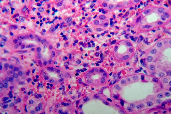
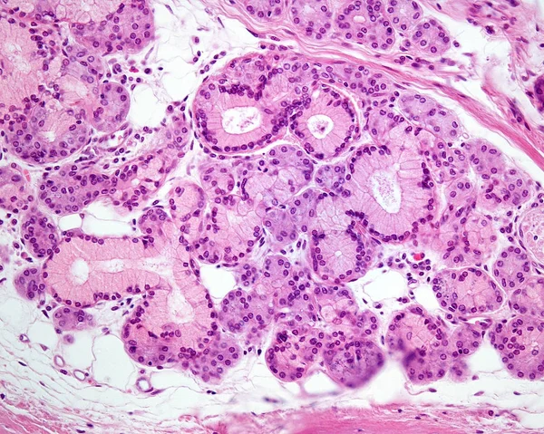
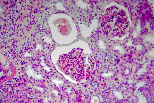
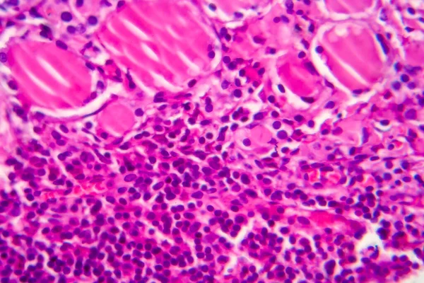
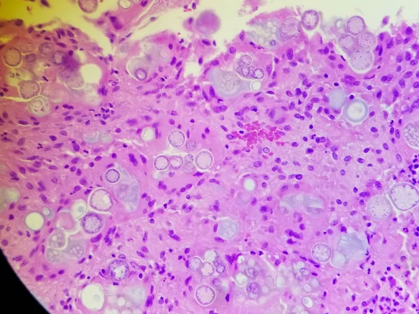
Stock image Coccidioides imitis spherules on H and E pathology specimen
Published: Dec.21, 2021 08:57:24
Author: CreativeEndeavors6@gmail.com
Views: 2
Downloads: 0
File type: image / jpg
File size: 2.13 MB
Orginal size: 2880 x 2160 px
Available sizes:
Level: bronze