Home
Images
Vectors
Videos
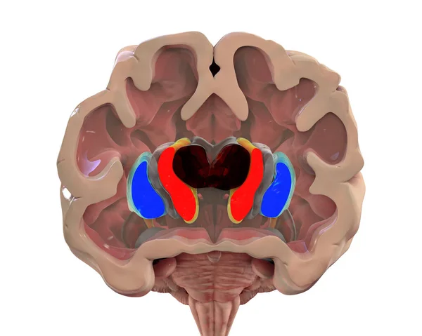
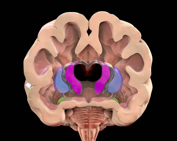
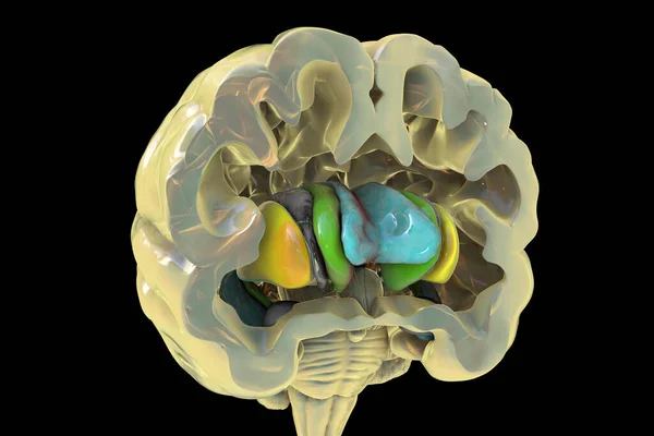
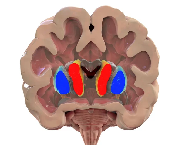
Stock image Coronal section of a healthy brain showing normal anatomy of basal baglia and ventricles, 3D illustration. Non-labelled version of the image
Published:
Jul.19, 2021 08:47:03
Author:
katerynakon
Views:
11
Downloads:
0
File type:
image
/
jpg
File size:
6.86 MB
Orginal size:
6250
x
5000
px
Available sizes:
S
(500 x 400)
M
(1000 x 800)
L
(2000 x 1600)
XL
(6250 x 5000)
EL0
(6250 x 5000)
Level:
silver
Download Image
Image topics:
White
Illustration
Isolated
Human
Healthy
Head
Medicine
Medical
3d
Body
Disease
Intelligence
Science
Section
Organ
Control
Cortex
Mind
Motor
Matter
Nucleus
Anatomy
Normal
Brain
Central
Nervous
Disorder
Neurology
Corpus
Anatomical
Dorsal
Coronal
Associative
Nuclei
Putamen
Neuro
Basal
Claustrum
Inhibitory
Parkinson
Black Background
Caudate
Ganglia
Striatum
Grey Matter
Lateral Ventricle
Corpus Callosum
Caudate Nucleus
Globus Pallidum
Similar stock images
Coronal Section Of A Brain Of A Person With Huntington's Disease Showing Enlarged Anterior Horns Of The Lateral Ventricles, Degeneration And Atrophy Of The Dorsal Striatum, 3D Illustration
6250 × 5000
Coronal Section Of A Brain Of A Person With Huntington's Disease Showing Enlarged Anterior Horns Of The Lateral Ventricles, Degeneration And Atrophy Of The Dorsal Striatum, 3D Illustration
5180 × 4144
3D Rendering Illustration Brown Organic Human Brain Side View Isolated On Pastel Blue Background With Separation Clipping Path Of Main Section
7200 × 4680
Human Brain Lobes Anatomy For Medical Concept 3D Illustration
4096 × 4096
Human Brain Anatomy, Basal Ganglia. 3D Illustration Showing Caudate Nucleus (green), Putamen (yellow), And Lateral Ventricles (blue)
6000 × 4000