
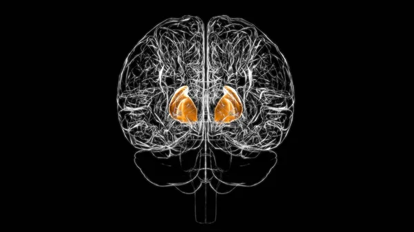
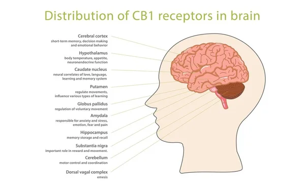
Stock image Caudate Nucleus
3d Rendered Medically Accurate Illustration Of A Young Girl Brains Anatomy-the Caudate Nucleus
Image, 7.67MB, 4000 × 4200 jpg

OCD Obsessive Compulsive Disorder Labeled Explanation Vector Illustration.
Vector, 9.26MB, 4000 × 4000 eps
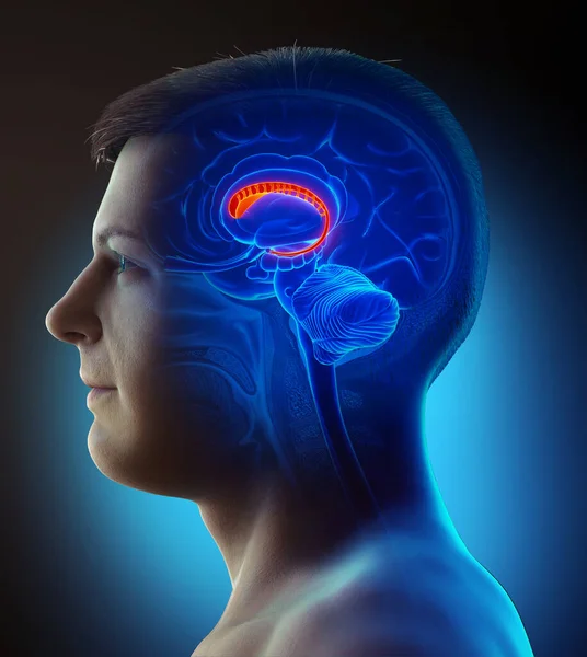
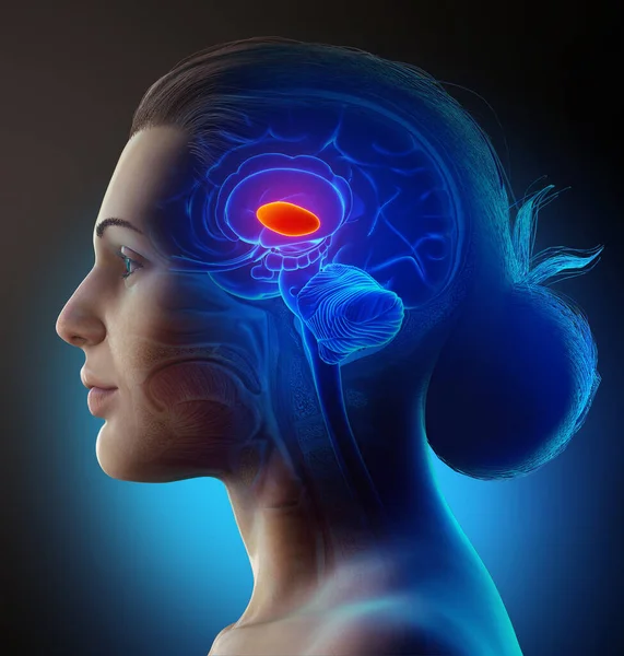
3d Rendered Medically Accurate Illustration Of A Male Brains Anatomy-the Caudate Nucleus
Image, 3.75MB, 3758 × 4200 jpg
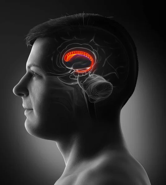
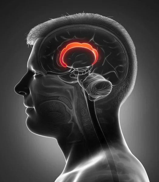
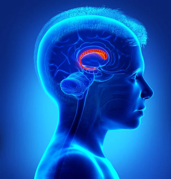
3d Rendered Medically Accurate Illustration Of A Male Brains Anatomy-the Caudate Nucleus
Image, 3.9MB, 3669 × 4200 jpg
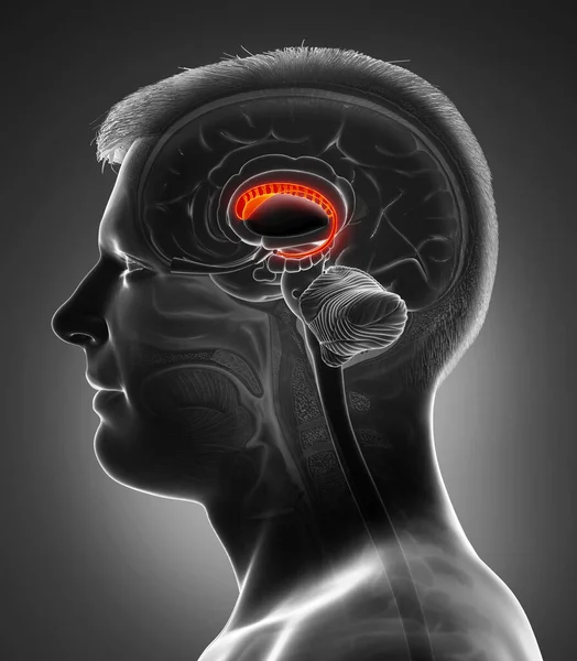
3d Rendered Medically Accurate Illustration Of A Male Brains Anatomy-the Caudate Nucleus
Image, 5.44MB, 3758 × 4200 jpg

Coronal Section Of A Healthy Brain Showing Normal Anatomy Of Basal Baglia And Ventricles, 3D Illustration
Image, 4.88MB, 6000 × 4000 jpg
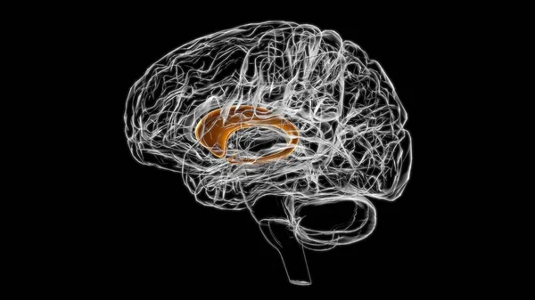
3d Rendered Medically Accurate Illustration Of A Female Brain Anatomy- The Thalamus
Image, 6.81MB, 4000 × 4200 jpg


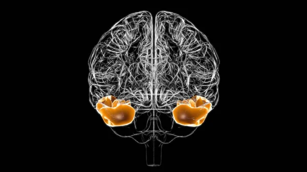
3d Rendered Medically Accurate Illustration Of A Female Brain Anatomy-the Lateral Globus Pallidus
Image, 6.8MB, 4000 × 4200 jpg
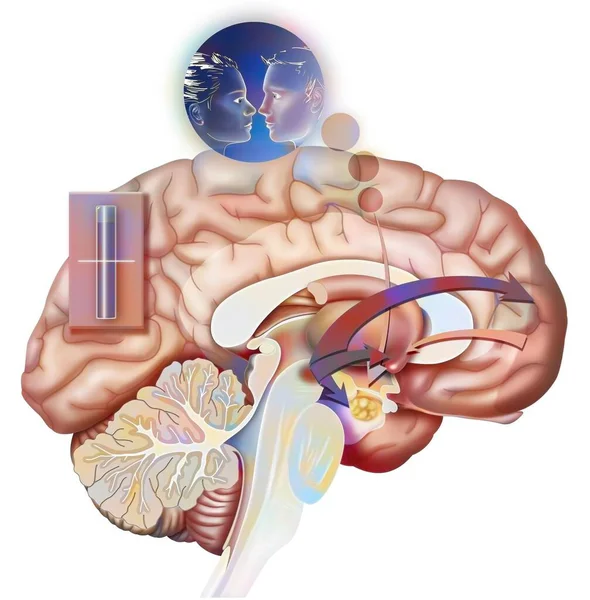
Desire And Rupture (state 2): Reaction Of The Brain When One Is In Love.
Image, 1.42MB, 4016 × 4085 jpg
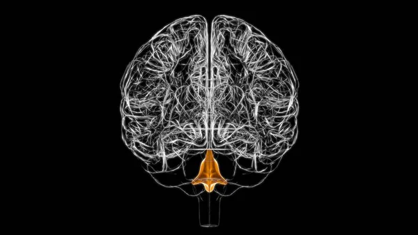
Brain Fourth Ventricles Of The Brain Anatomy For Medical Concept 3D Illustration
Image, 1.82MB, 3840 × 2160 jpg
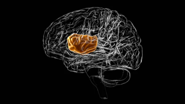
Brain Inferior Temporal Gyrus Anatomy For Medical Concept 3D Illustration
Image, 1.79MB, 3840 × 2160 jpg

Brain Inferior Temporal Gyrus Anatomy For Medical Concept 3D Illustration
Image, 1.73MB, 3840 × 2160 jpg

3d Rendered Medically Accurate Illustration Of A Male Brain Anatomy-the Lateral Globus Pallidus
Image, 3.86MB, 3669 × 4200 jpg

Brain Inferior Frontal Gyrus Anatomy For Medical Concept 3D Illustration
Image, 1.78MB, 3840 × 2160 jpg
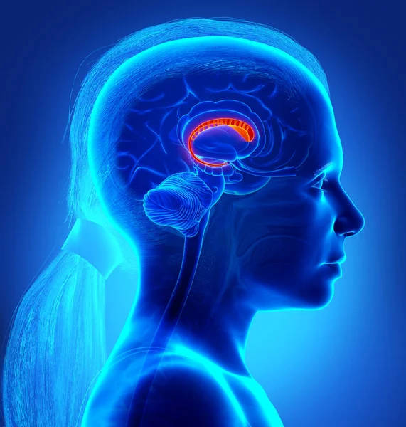
3d Rendered Medically Accurate Illustration Of A Young Boy Brains Anatomy-the Caudate Nucleus
Image, 6MB, 4000 × 4200 jpg

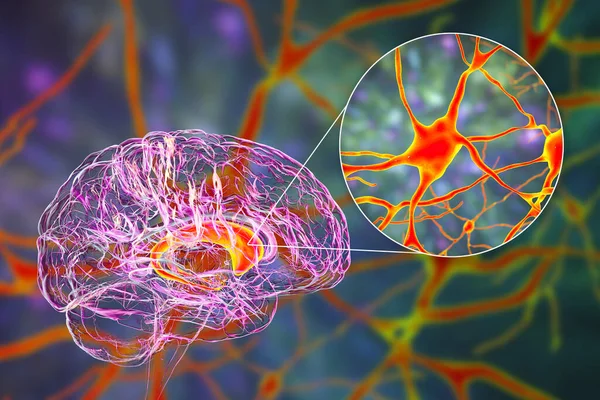
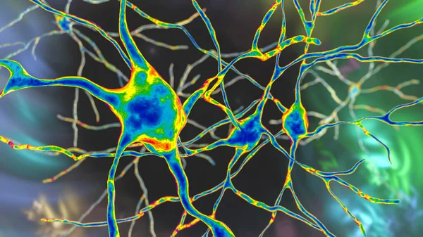
Caudate Nuclei In Human Brain And Its Neurons, 3D Illustration. The Caudate Nucleus Is A Component Of The Basal Ganglia, It Plays Role In Choreas, Neurodegenerative And Other Brain Diseases
Image, 16.77MB, 8157 × 5438 jpg
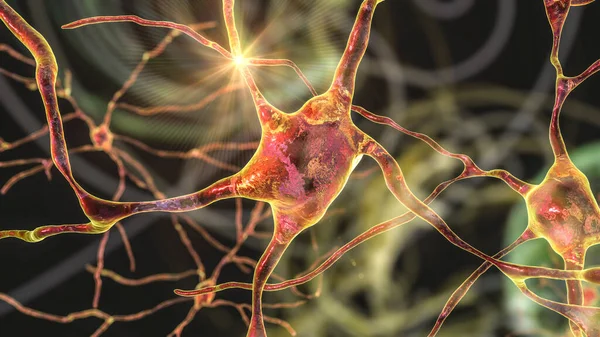
Neurons Of Dorsal Striatum, 3D Illustration. The Dorsal Striatum Is A Nucleus In The Basal Ganglia, Degrading Of Its Neurons Plays A Crucial Role In The Development Of Huntington's Disease
Image, 12.53MB, 7200 × 4050 jpg

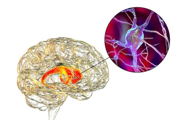
Anti-basal Ganglia Antibodies. 3D Conceptual Illustration Showing Molecules Of Immunoglobulins Attacking Dorsal Striatum Highlighted In The Human Brain. They Are Found In Post-rheumatic Fever Chorea
Image, 21.21MB, 10147 × 6765 jpg
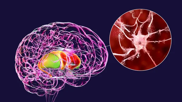
Dorsal Striatum Highlighted In Human Brain And Close-up View Of Degrading Neurons Of Dorsal Striatum Seen In Huntington's Disease, 3D Illustration
Image, 16.37MB, 8672 × 4878 jpg

Caudate Nuclei In Human Brain And Its Neurons, 3D Illustration. The Caudate Nucleus Is A Component Of The Basal Ganglia, It Plays Role In Choreas, Neurodegenerative And Other Brain Diseases
Image, 12.8MB, 8157 × 5438 jpg
Neurons Of Dorsal Striatum, 3D Illustration. The Dorsal Striatum Is A Nucleus In The Basal Ganglia, Degrading Of Its Neurons Plays A Crucial Role In The Development Of Huntington's Disease
Image, 8.02MB, 7200 × 4050 jpg

Destruction Of Neurons Of The Caudate Nucleus, Conceptual 3D Illustration. Caudate Nucleus Belongs To The Brain Basal Ganglia, Its Neurons Are Damaged In Huntingon's Disease And Other Choreas
Image, 16.49MB, 7996 × 5331 jpg
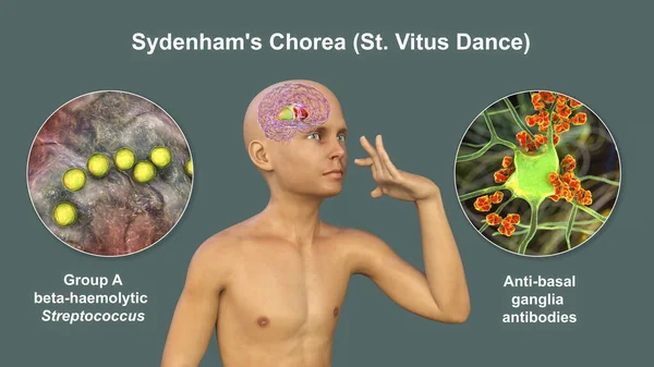
Sydenham's Chorea, An Autoimmune Disease That Results From Streptococcus Infection, Formation Of Anti-neuronal Antibodies Damaging Brain Basal Ganglia That Cause Involuntary Movements, 3D Illustration
Image, 16.36MB, 8737 × 4914 jpg
Caudate Nuclei Highlighted In Human Brain, 3D Illustration. The Caudate Nucleus Is A Component Of The Basal Ganglia, It Is Associated With Motor Processes And Plays Role In Parkinson's Disease
Image, 9.68MB, 6000 × 4000 jpg
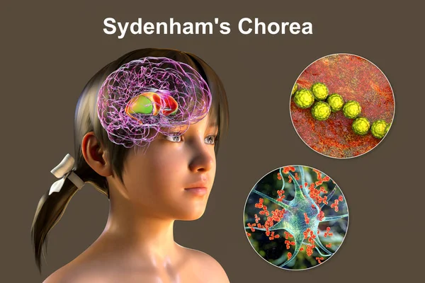
Sydenham's Chorea, An Autoimmune Disease That Results From Streptococcus Infection, Formation Of Anti-neuronal Antibodies Damaging Brain Basal Ganglia That Cause Involuntary Movements, 3D Illustration
Image, 39.11MB, 10609 × 7073 jpg
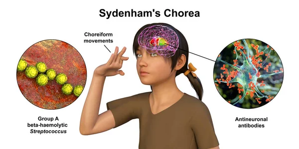
Sydenham's Chorea, An Autoimmune Disease That Results From Streptococcus Infection, Formation Of Anti-neuronal Antibodies Damaging Brain Basal Ganglia That Cause Involuntary Movements, 3D Illustration
Image, 12.54MB, 8488 × 4244 jpg
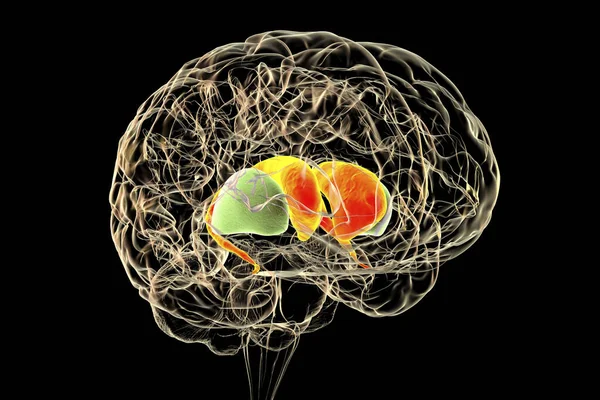
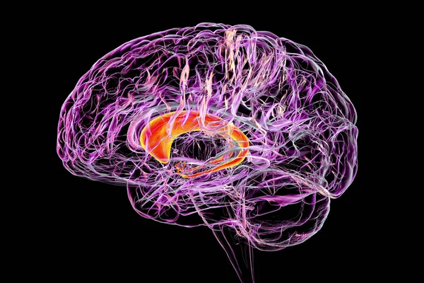
Dorsal Striatum In The Human Brain, 3D Illustration. It Is A Nucleus In The Basal Ganglia, Consists Of The Caudate Nucleus (red) And The Putamen (green), Is A Component Of The Motor And Reward Systems
Image, 6.21MB, 6000 × 4000 jpg
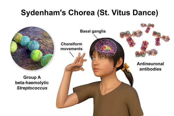
Sydenham's Chorea, An Autoimmune Disease That Results From Streptococcus Infection, Formation Of Anti-neuronal Antibodies Damaging Brain Basal Ganglia That Cause Involuntary Movements, 3D Illustration
Image, 10.04MB, 7883 × 5255 jpg

Dorsal Striatum Highlighted In Child's Brain And Close-up View Of Its Neurons, 3D Illustration. It Is A Nucleus In The Basal Ganglia, A Component Of The Motor And Reward Systems
Image, 23.52MB, 9339 × 6226 jpg

Neurons Of Dorsal Striatum, 3D Illustration. Dorsal Striatum Is A Nucleus In The Basal Ganglia, Degrading Of Its Neurons Plays Crucial Role In Development Of Huntington's Disease
Image, 8.32MB, 7200 × 4050 jpg
Caudate Nuclei In Human Brain And Its Neurons, 3D Illustration. The Caudate Nucleus Is A Component Of The Basal Ganglia, It Plays Role In Choreas, Neurodegenerative And Other Brain Diseases
Image, 11.51MB, 7996 × 5331 jpg
Neurons Of Dorsal Striatum, 3D Illustration. Dorsal Striatum Is A Nucleus In The Basal Ganglia, Degrading Of Its Neurons Plays Crucial Role In Development Of Huntington's Disease
Image, 12.86MB, 7200 × 4050 jpg

Brain Dorsal Striatum Anatomy, 3D Illustration. The Dorsal Striatum Consists Of The Caudate Nucleus (orange) And The Putamen (blue). Amygdala Is Colored In Red. Front View
Image, 5.28MB, 6000 × 4000 jpg


Dorsal Striatum Highlighted In Child's Brain And Close-up View Of Its Neurons, 3D Illustration. It Is A Nucleus In The Basal Ganglia, A Component Of The Motor And Reward Systems
Image, 31.18MB, 9339 × 6226 jpg
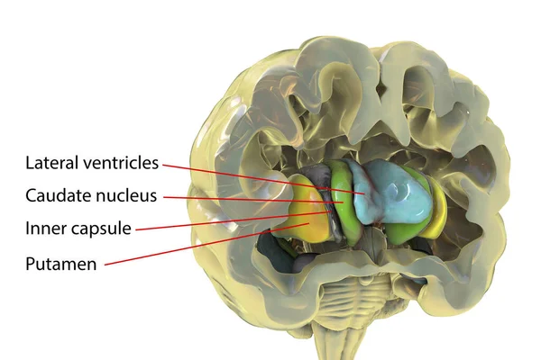
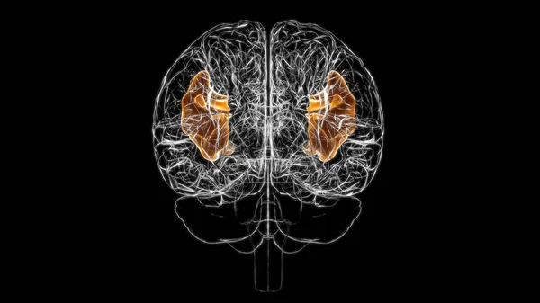
Human Brain Anatomy, Basal Ganglia. 3D Illustration Showing Caudate Nucleus (green), Putamen (yellow), And Lateral Ventricles (blue)
Image, 6.25MB, 6252 × 4168 jpg
Page 1 >> Next