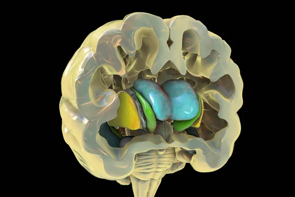
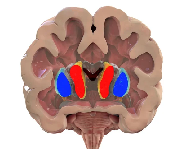
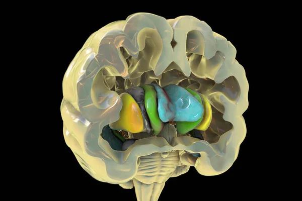
Stock image Human brain anatomy, basal ganglia. 3D illustration showing caudate nucleus (green), putamen (yellow), and lateral ventricles (blue)
Published: May.24, 2021 13:03:01
Author: katerynakon
Views: 6
Downloads: 0
File type: image / jpg
File size: 5.95 MB
Orginal size: 6000 x 4000 px
Available sizes:
Level: silver