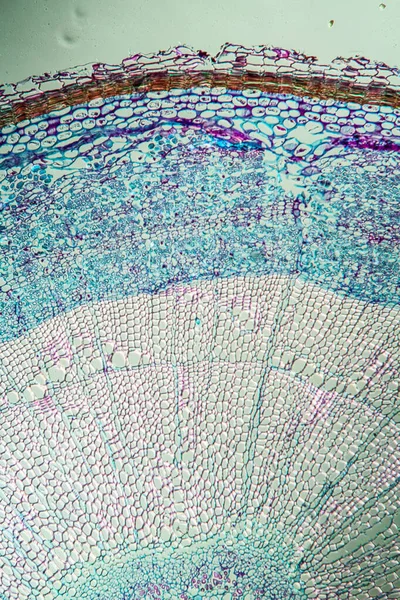
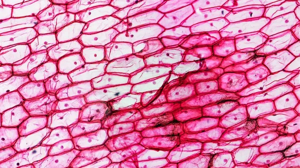
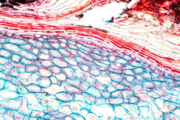
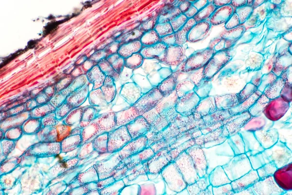
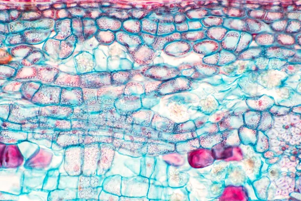
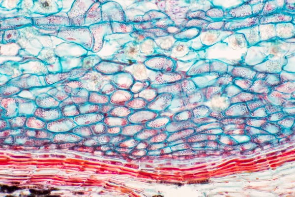
Stock image Cross sections of plant stem under microscope view show Structure of Collenchyma Cells for education botany.

Published: Aug.25, 2020 15:43:52
Author: tonaquatic19
Views: 1
Downloads: 0
File type: image / jpg
File size: 17 MB
Orginal size: 6000 x 4000 px
Available sizes:
Level: bronze
Similar stock images
Cross Sections Of Plant Stem Under Microscope View Show Structure Of Collenchyma Cells For Education Botany.
6000 × 4000
Cross Sections Of Plant Stem Under Microscope View Show Structure Of Collenchyma Cells For Education Botany.
6000 × 4000
Cross Sections Of Plant Stem Under Microscope View Show Structure Of Collenchyma Cells For Education Botany.
6000 × 4000
Cross Sections Of Plant Stem Under Microscope View Show Structure Of Collenchyma Cells For Education Botany.
6000 × 4000