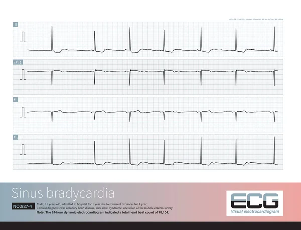
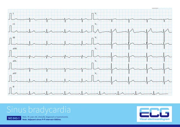
Stock image Generally, when the sinus heart rate is below 60 beats per minute, it is called sinus bradycardia. This arrhythmia can be both physiological and often pathological.
Published: Mar.21, 2023 08:44:50
Author: asia11m
Views: 18
Downloads: 1
File type: image / jpg
File size: 20.38 MB
Orginal size: 10000 x 7069 px
Available sizes:
Level: beginner