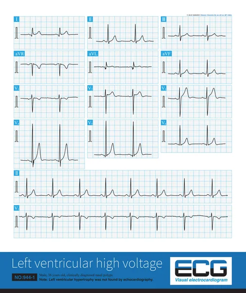
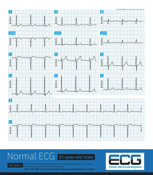
Stock image Note that the V3 lead of this ECG shows that the amplitude of R wave is greater than the amplitude of S wave, and there is counterclockwise rotation.
Published: Mar.06, 2023 10:29:20
Author: asia11m
Views: 22
Downloads: 1
File type: image / jpg
File size: 33.74 MB
Orginal size: 10000 x 11469 px
Available sizes:
Level: beginner