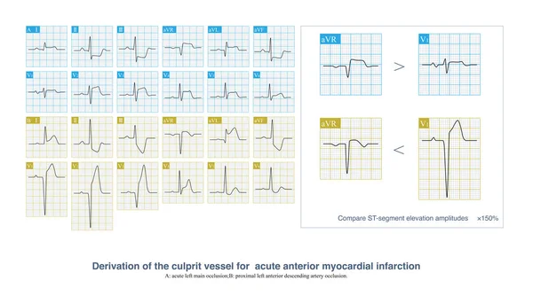
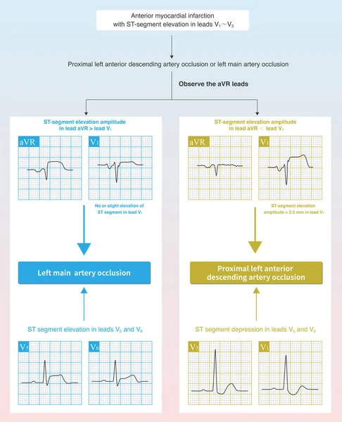
Stock image In case of acute anterior myocardial infarction, the characteristics of ST segment elevation in ECG can be used to deduce whether the culprit vessel system is the left main trunk or the proximal LAD.
Published: Sep.22, 2022 07:37:15
Author: asia11m
Views: 23
Downloads: 1
File type: image / jpg
File size: 17.81 MB
Orginal size: 10000 x 12321 px
Available sizes:
Level: beginner