Home
Images
Vectors
Videos
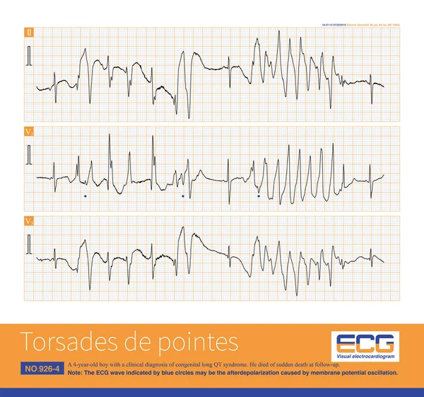
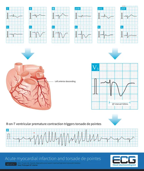
Stock image Male, 75 years old, clinically diagnosed as acute anterior septal and high lateral myocardial infarction.The culprit vessel was located in the LAD proximal segment.Prolonged QT interval with TDP.
Published:
Jun.10, 2022 09:12:51
Author:
asia11m
Views:
71
Downloads:
0
File type:
image
/
jpg
File size:
22.45 MB
Orginal size:
10000
x
11911
px
Available sizes:
S
(420 x 500)
M
(840 x 1000)
L
(1679 x 2000)
XL
(10000 x 11911)
EL0
(10000 x 11911)
Level:
beginner
Download Image
Image topics:
White
Background
Graphic
Illustration
Paper
Human
Health
Healthy
Medicine
Medical
Care
Technology
Line
Cardiology
Heart
Hospital
Monitor
Pulse
Test
Disease
Education
Science
Death
Analysis
Graph
Rhythm
Diagnosis
Heartbeat
Artery
Cardiogram
Cardiac
Ekg
Electrocardiogram
Ecg
Chest Pain
Myocardial Infarction
Coronary Heart Disease
Coronary Artery
Sudden Death
Myocardial Ischemia
Limb Leads
Acute Coronary Syndrome
St Segment
Ecg Education
Qrs Wave
U Wave
T Wave Inversion
Ischemic Chest Pain
Long Qt
Similar stock images
A 4-year-old Boy With A Clinical Diagnosis Of Long QT Syndrome. No Genetic Testing Was Done During Hospitalization. The Child Died Suddenly During Follow-up.
10000 × 9356
When The Frequency Of Sinus Impulses Exceeds 100 Beats Per Minute, Called Sinus Tachycardia, It Is A Common Clinical Arrhythmia. Treatment Focuses On Finding The Cause Of The Increased Heart Rate.
10000 × 7070
Torsade De Pointes Refers To The Pleomorphic Ventricular Tachycardia That Occurs In The Background Of Long QT Interval, And The Polarity Of QRS Wave Twists Around The Equipotential Line.
10000 × 5808
Normal Electrocardiogram Of A 34 Year Old Chinese Healthy Woman.The Rhythm Was Sinus Rhythm Without Other Abnormal ECG Changes.
4000 × 5237
The Amplitude Of T Wave In Lead V1 Is Most Commonly Seen In Hypertension And Coronary Heart Disease, Followed By Other Cardiovascular Diseases, And Also Seen In Normal Individuals.
10000 × 6534
The U-wave Is A Low Amplitude Electrocardiogram That Follows The T-wave, Usually With An Amplitude Of No More Than 3.3mm. The Maximum Amplitude Is Commonly Seen In The V2-V4 Lead.
10000 × 7706
Cardiac Surgery Is One Of The Common Causes Of Iatrogenic Atrioventricular Block. Once Atrial Fibrillation Exhibits A Slow And Regular Ventricular Rhythm, It Is Important To Be Alert To 3 AVB.
10000 × 11558
Male, 75 Years Old, Clinically Diagnosed As Acute Anterior Septal Myocardial Infarction, Prolonged QT Interval, Complicated With Torsade De Pointes.
14017 × 6675