Home
Images
Vectors
Videos
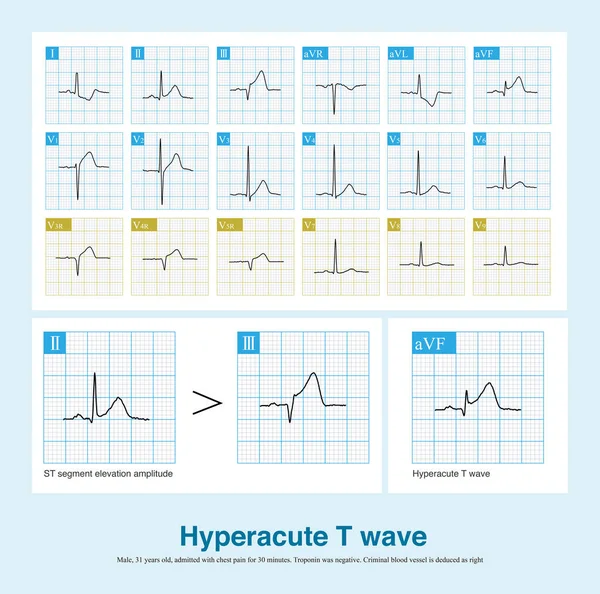
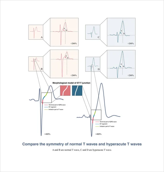
Stock image It is worth noting that many electrocardiographic textbooks believe that hyperacute T waves are symmetrical. In fact, they are not symmetrical.
Published:
Dec.05, 2022 09:02:30
Author:
asia11m
Views:
38
Downloads:
0
File type:
image
/
jpg
File size:
9.23 MB
Orginal size:
10000
x
10486
px
Available sizes:
S
(477 x 500)
M
(954 x 1000)
L
(1907 x 2000)
XL
(10000 x 10486)
EL0
(10000 x 10486)
Level:
beginner
Download Image
Image topics:
Blue
Background
Graphic
Illustration
Rate
Medical
Line
Wave
Cardiology
Heart
Pulse
Disease
Grid
Science
Death
Analysis
Graph
Rhythm
Heartbeat
Artery
Cardiogram
Cardiac
Ekg
Electrocardiogram
Ecg
Thrombosis
Heart Disease
Chest Pain
Myocardial Infarction
Coronary Heart Disease
Coronary Artery
Ecg Graph
Troponin
Sudden Death
Ischemic Heart Disease
Myocardial Ischemia
Acute Coronary Syndrome
Coronary Spasm
St Segment
Ecg Education
Qrs Wave
Chest Lead
St Segment Elevation
St Segment Depression
Left Main Artery
Left Main Occlusion
Hyperacute T Wave
Early Repolarization
Similar stock images
Female, 64 Years Old, With A History Of Hypertension And Hyperlipidemia. Repeated Chest Pain In The Past 6 Months. CA Confirmed Spasm Of The LAD, And No Stenosis In All Three Coronary Arteries.
10000 × 8293
In The Early Stage Of Acute Myocardial Infarction, T Wave Is Upright, With Increased Amplitude And Symmetry, With Or Without ST Segment Elevation, Which Is Called Hyperacute T Wave.
10000 × 9910
In Case Of Acute Anterior Myocardial Infarction, The Characteristics Of ST Segment Elevation In ECG Can Be Used To Deduce Whether The Culprit Vessel System Is The Left Main Trunk Or The Proximal LAD.
10000 × 12321
If Atrioventricular Dissociation And Ventricular Capture Are Found During Wide QRS Tachycardia, It Supports The Diagnosis Of Ventricular Tachycardia With High Specificity And Poor Sensitivity.
10000 × 3269
Male, 58 Years Old, Admitted With Chest Pain For 30 Minutes. ECG Showed Hyperacute T Wave, Acute Myocardial Ischemia Involving High Lateral Wall And Anterior Wall. The Disappearance Of S Wave In Leads V3 And V4 Was Grade III Myocardial Ischemia.
10000 × 7384
In Acute Extensive Anterior Myocardial Infarction, The Culprit Vessel May Be Occlusion Of The Left Main Artery Or Proximal Left Anterior Descending Artery, Either With A High Risk Of Death.
10000 × 5719
Sometimes, Because The QRS Axis Is In The Upper Left Quadrant, The High-amplitude R Wave Of Left Ventricular Hypertrophy Occurs In The Limb Leads, And Left Chest Leads Is Normal.
10000 × 10111