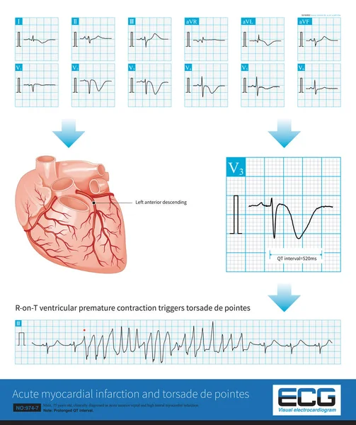
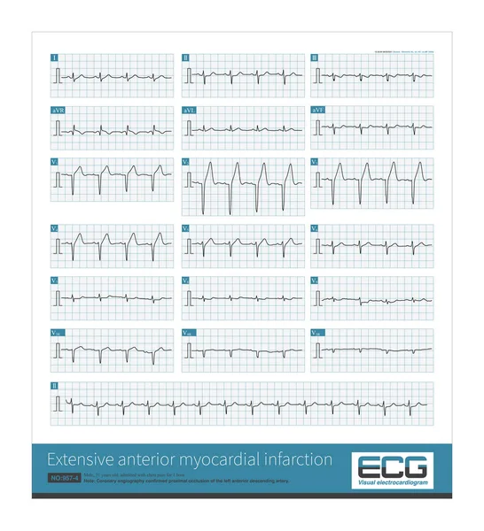
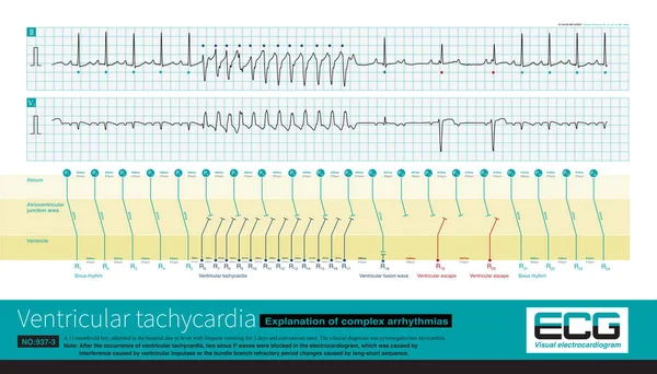
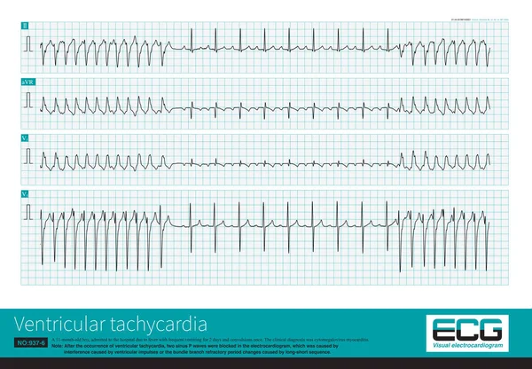
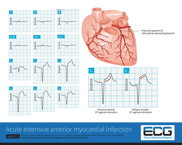
Stock image Male, 64 years old, admitted with chest pain for 4 hours. The clinical diagnosis was extensive anterior and inferior myocardial infarction. The culprit vessel was the left anterior descending branch.
Published: Jun.27, 2022 08:21:12
Author: asia11m
Views: 62
Downloads: 0
File type: image / jpg
File size: 11.63 MB
Orginal size: 10000 x 7987 px
Available sizes:
Level: beginner