Home
Images
Vectors
Videos
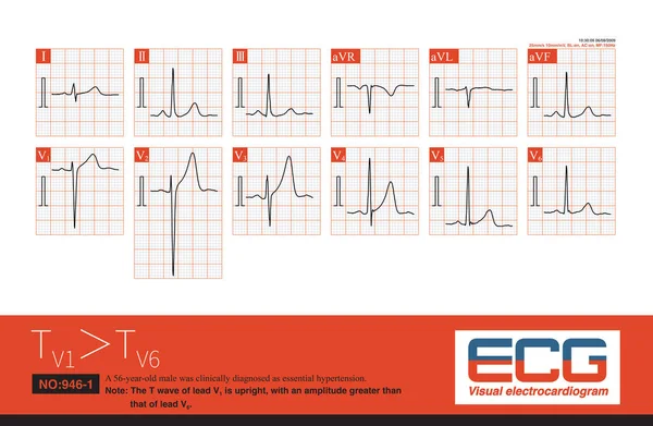
Stock image The amplitude of T wave in lead V1 is most commonly seen in hypertension and coronary heart disease, followed by other cardiovascular diseases, and also seen in normal individuals.
Published:
Apr.04, 2023 09:27:22
Author:
asia11m
Views:
11
Downloads:
0
File type:
image
/
jpg
File size:
11.95 MB
Orginal size:
10000
x
6534
px
Available sizes:
S
(500 x 327)
M
(1000 x 653)
L
(2000 x 1307)
XL
(10000 x 6534)
EL0
(10000 x 6534)
Level:
beginner
Download Image
Image topics:
Graphic
Illustration
Human
Health
Healthy
Medicine
Healthcare
Medical
Wave
Cardiology
Heart
Hospital
Pulse
Body
Disease
Education
Science
Death
Organ
Graph
Beat
Rhythm
Diagnosis
Hypertension
Amplitude
Cardiogram
Cardiac
Ekg
Electrocardiogram
Atrium
Ecg
Arrhythmia
Arrhythmias
Heart Disease
Repolarization
Cardiac Arrest
Cardiomyopathy
Coronary Heart Disease
Left Ventricle
Coronary Artery Disease
Sudden Death
ECG Monitoring
Left Ventricular Hypertrophy
Ecg Education
Ventricular Arrhythmia
Diagonal Artery
Ventricular Repolarization
Escape Rhythm
Similar stock images
Cardiac Surgery Is One Of The Common Causes Of Iatrogenic Atrioventricular Block. Once Atrial Fibrillation Exhibits A Slow And Regular Ventricular Rhythm, It Is Important To Be Alert To 3 AVB.
10000 × 11558
Normal Electrocardiogram Of A 34 Year Old Chinese Healthy Woman.The Rhythm Was Sinus Rhythm Without Other Abnormal ECG Changes.
4000 × 5237
Male, 75 Years Old, Clinically Diagnosed As Acute Anterior Septal And High Lateral Myocardial Infarction.The Culprit Vessel Was Located In The LAD Proximal Segment.Prolonged QT Interval With TDP.
10000 × 11911
The U-wave Is A Low Amplitude Electrocardiogram That Follows The T-wave, Usually With An Amplitude Of No More Than 3.3mm. The Maximum Amplitude Is Commonly Seen In The V2-V4 Lead.
10000 × 7706
When The Frequency Of Sinus Impulses Exceeds 100 Beats Per Minute, Called Sinus Tachycardia, It Is A Common Clinical Arrhythmia. Treatment Focuses On Finding The Cause Of The Increased Heart Rate.
10000 × 7070
In The Same Patient, T Wave Notch Can Be Manifested As T Wave Descending Branch Notch, Double-peak And Flat-peak T Wave.
10000 × 8870
A 4-year-old Boy With A Clinical Diagnosis Of Long QT Syndrome. No Genetic Testing Was Done During Hospitalization. The Child Died Suddenly During Follow-up.
10000 × 9356
A 36 Year Old Man Survived CPR After Sudden Syncope. The Electrocardiogram Was Suggestive Of Brugada Syndrome Type 1. Implantation Of ICD Therapy.
9000 × 13286