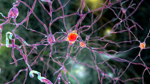
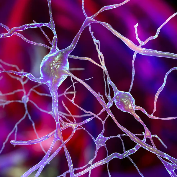
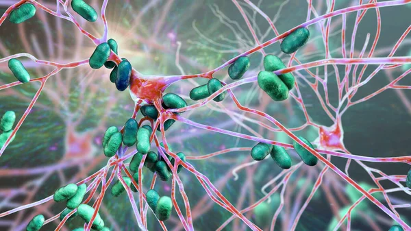
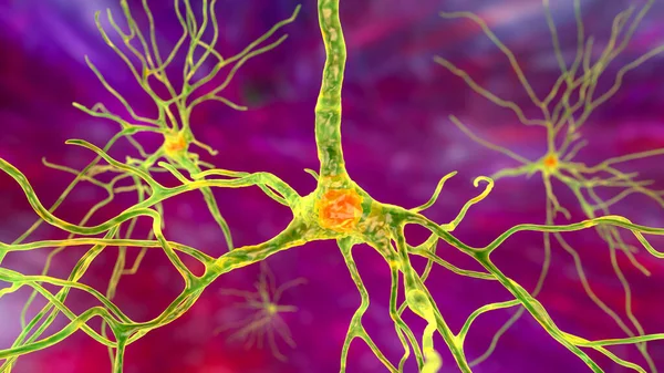
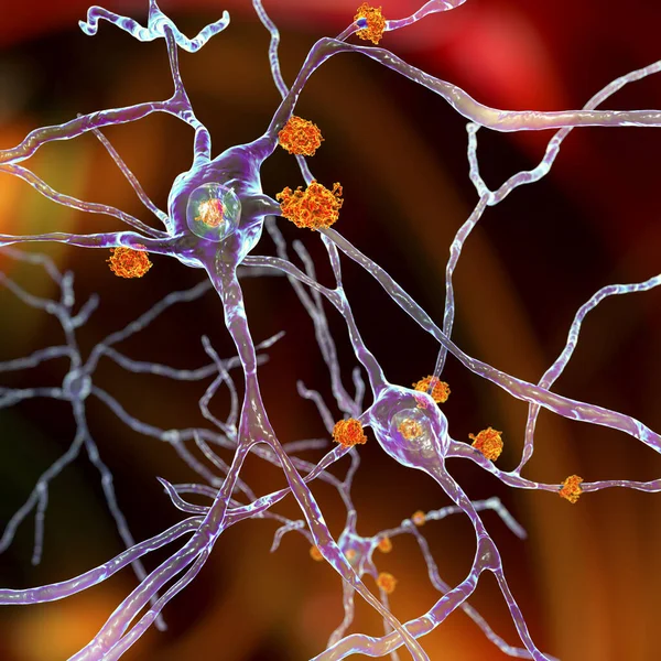
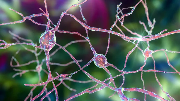
Stock image Neurons of Dorsal striatum, 3D illustration. The dorsal striatum is a nucleus in the basal ganglia, degrading of its neurons plays a crucial role in the development of Huntington's disease
Published: Jun.18, 2021 08:44:25
Author: katerynakon
Views: 3
Downloads: 0
File type: image / jpg
File size: 10.06 MB
Orginal size: 7200 x 4050 px
Available sizes:
Level: silver