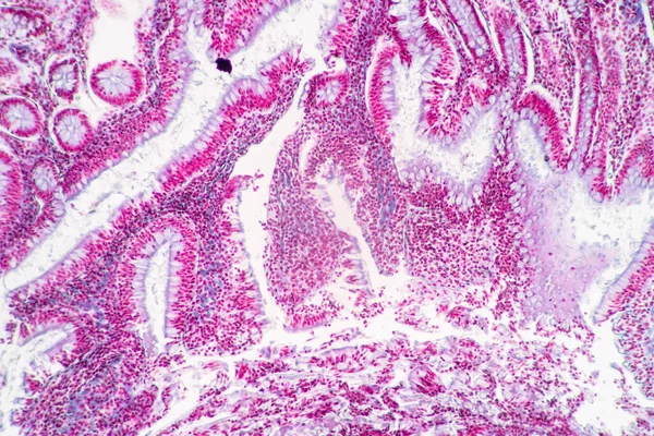
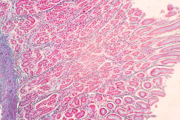
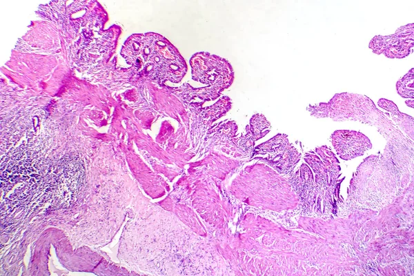
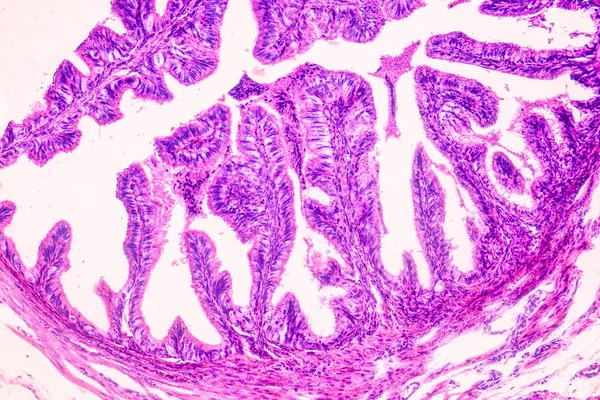
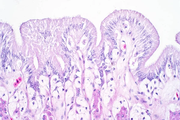
Stock image Ovarian mucinous cystadenoma (15-20% of ovarian tumours) is a usually large and benign tumour showing glands and cysts, with filiform papillae with fibrovascular cores, lined by benign columnar epithelium.

Published: Jul.04, 2022 16:30:27
Author: jlcalvo@ucm.es
Views: 5
Downloads: 0
File type: image / jpg
File size: 10.54 MB
Orginal size: 3840 x 3072 px
Available sizes:
Level: beginner