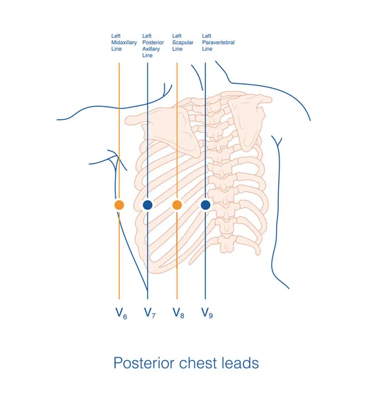
Stock image Sometimes, it is necessary to place electrodes in the high intercostal space at the conventional chest lead electrode placement position, which is called a high intercostal chest lead.

Published: Jul.27, 2023 13:45:03
Author: asia11m
Views: 22
Downloads: 1
File type: image / jpg
File size: 10.05 MB
Orginal size: 10000 x 10000 px
Available sizes:
Level: beginner