Home
Images
Vectors
Videos
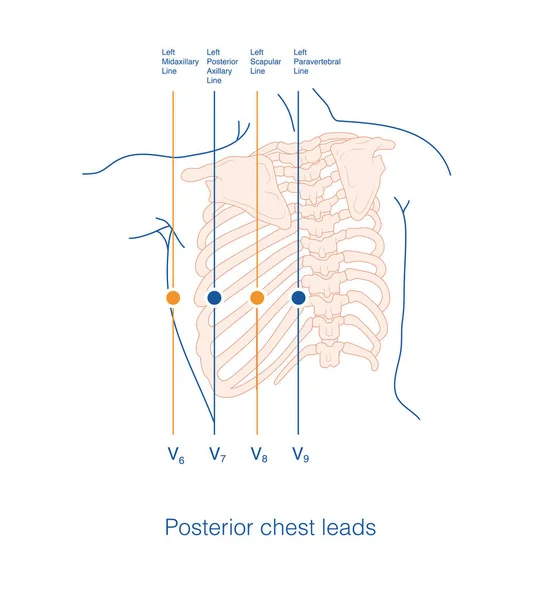
Stock image The chest electrode of ECG includes conventional chest lead, posterior wall lead and right ventricular lead. The placement of chest lead electrode shall comply with the specification.
Published:
Jun.14, 2022 18:42:42
Author:
asia11m
Views:
173
Downloads:
0
File type:
image
/
jpg
File size:
4.93 MB
Orginal size:
5512
x
5511
px
Available sizes:
S
(500 x 500)
M
(1000 x 1000)
L
(2000 x 2000)
XL
(5512 x 5511)
EL0
(5512 x 5511)
Level:
beginner
Download Image
Image topics:
White
Background
Graphic
Illustration
Paper
Isolated
Human
Health
Healthy
Medicine
Medical
Care
Line
Wave
Structure
Cardiology
Heart
Monitor
Pulse
Body
Disease
Education
Science
Death
Organ
Lead
Analysis
Graph
Scientific
Rhythm
Heartbeat
Anatomy
Cardiogram
Cardiac
Ekg
Electrocardiogram
Ecg
Anatomical
Arrhythmia
Heart Disease
Chest Pain
Internal Medicine
Medical Illustration
Sudden Death
Limb Leads
St Segment
Ecg Education
Qrs Wave
Chest Lead
Similar stock images
Place The Surface Electrocardiogram Exploration Electrode At The Specified Anatomical Position On The Left Back To Form A Posterior Lead Consisting Of V7-V9 Leads.
10000 × 11258
When A Single Limb ECG Is Probed, The Probe Lead Is Removed From The Central Terminal, Which Is Formed By The Connection Of The Remaining Two Leads.
10000 × 10000
Chest Lead Is A Lead System Used To Detect The Transverse Electrical Activity Of The Heart, Typically Consisting Of Six Electrodes Placed In The Right And Front Of The Left Chest.
10000 × 10000
Illustration Of Positional Structure And Overlap Of Abdominal Muscle Groups Illustration Frontal And Cross-sectional Views
4500 × 3938
In Case Of Acute Anterior Myocardial Infarction, The Characteristics Of ST Segment Elevation In ECG Can Be Used To Deduce Whether The Culprit Vessel System Is The Left Main Trunk Or The Proximal LAD.
10000 × 12321
Airways Structure
5000 × 5000
Stress Responce System
4168 × 4168
Stress Responce System
4167 × 4168