Home
Images
Vectors
Videos
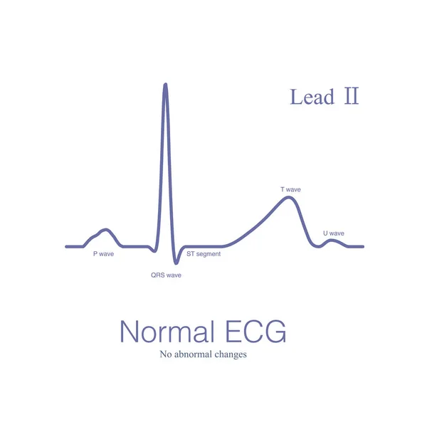
Stock image Sometimes, there may be slight non-specific changes and normal variations in the electrocardiogram, which are often due to physiological reasons and have no clinical therapeutic significance.
Published:
Feb.24, 2024 13:54:28
Author:
asia11m
Views:
1
Downloads:
1
File type:
image
/
jpg
File size:
13.15 MB
Orginal size:
10000
x
11438
px
Available sizes:
S
(437 x 500)
M
(874 x 1000)
L
(1749 x 2000)
XL
(10000 x 11438)
EL0
(10000 x 11438)
Level:
beginner
Download Image
Image topics:
Illustration
Human
Health
Medical
Cardiology
Heart
Hospital
Disease
Education
Death
Graph
Beat
Rhythm
Diagnosis
Heartbeat
Anatomy
Cardiac
Ekg
Electrocardiogram
Ecg
Arrhythmia
Heart Disease
Cardiac Arrest
Human Organs
Sudden Death
Conduction System
ECG Monitoring
Atrioventricular Node
Normal Variant
Sinus Node
P Wave
QRS Complex
St Segment
Pr Segment
Pr Interval
Ecg Education
Qrs Wave
U Wave
Chest Leads
His Bundle
J Point
Right Bundle Branch
Left Bundle Branch
Bachmann Bundle
Terminal Purkinje Fiber Mesh
Internodal Conduction Pathway
Otherwise Normal Ecg
Roughly Normal
Similar stock images
A Normal ECG Is An ECG That Does Not Have Any Abnormal Changes And Can Be Seen In Healthy People As Well As In People With Heart Disease.
10000 × 11438
A Normal ECG Is An ECG That Does Not Have Any Abnormal Changes And Can Be Seen In Healthy People As Well As In People With Heart Disease.
10000 × 5625
Sometimes, There May Be Slight Non-specific Changes And Normal Variations In The Electrocardiogram, Which Are Often Due To Physiological Reasons And Have No Clinical Therapeutic Significance.
10000 × 5625
When The Opening Of Left Anterior Descending Artery Is Occluded, It Can Cause A Large Area Of Extensive Anterior Myocardial Infarction (some Patients With Inferior Wall), And The Prognosis Is Poor.
10000 × 7063
A Normal ECG Is An ECG That Does Not Have Any Abnormal Changes And Can Be Seen In Healthy People As Well As In People With Heart Disease.
10000 × 10000
Common ECG Changes In Chronic Renal Insufficiency Include Left Ventricular Hypertrophy, Left Atrial Abnormality, Poor R Wave Progression And ST-T Changes.
10000 × 6551
Coronary Artery Spasm Causes Transmural Myocardial Ischemia, And ST Segment Elevation In ECG Has Localization Characteristics. Criminal Vessels Can Be Derived From ST Segment Elevation Leads In ECG.
10000 × 7684