Home
Images
Vectors
Videos
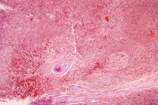
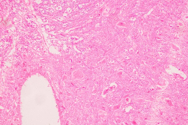
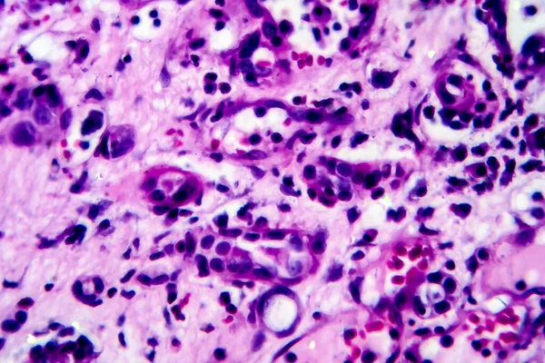
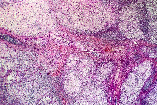
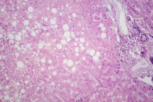
Stock image This is a pathological photo of a human gout nodule, showing the formation of pink amorphous eosinophilic substances by urate crystals.Magnify 40x.
Published:
May.11, 2024 10:58:35
Author:
asia11m
Views:
2
Downloads:
0
File type:
image
/
jpg
File size:
40.54 MB
Orginal size:
6000
x
6000
px
Available sizes:
S
(500 x 500)
M
(1000 x 1000)
L
(2000 x 2000)
XL
(6000 x 6000)
EL0
(6000 x 6000)
Level:
beginner
Download Image
Image topics:
Photography
Human
Light
Medicine
Illness
Medical
Heart
Cell
Disease
Cells
Congestion
Nucleus
Anatomy
Mucus
Acute
Deposit
Software
Tumor
Bleeding
Joints
Inflammation
Organs
Atrium
Infarction
Pathology
Tuberculosis
Arthritis
Staining
Serous
Nuclei
Biopsy
Atrophy
Glands
Tumors
Histology
Gout
Ischemia
Cytoplasm
Cross Section
Human Body
Fibrosis
Necrosis
Lymphocytes
Fibroblasts
Macrophages
Stroma
Intracellular
Red Blood Cells
Uric Acid
Neutrophils
Similar stock images
This Pathologic Photograph Highlights The Atrium Myxoma Cell Nest. Atrial Myxoma Is A Benign Tumor Of The Heart.
5500 × 7384
Almond Tonsils In Cross Section Under The Microscope 100x
5616 × 3744
Education Spinal Cord And Motor Neuron Under The Microscope In Lab.
6720 × 4480
Histopathology Of Interstitial Nephritis, Light Micrograph, Photo Under Microscope. High Magnification
4748 × 3165
Squamous Cell Carcinoma Diseased Tissue Under The Microscope 100x
5616 × 3744
Histopathology Of Portal Cirrhosis
4260 × 2840
Light Micrograph Revealing Liver Tissue With Fatty Infiltration, Indicative Of Hepatic Steatosis Or Fatty Liver Disease.
7178 × 4786
Human Prostate. Corpora Amylacea
4068 × 3072