Home
Images
Vectors
Videos
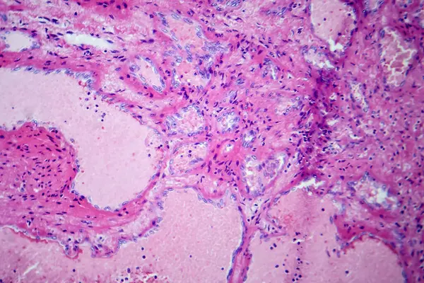
Stock image This photo shows the pink mucinous matrix of atrial myxoma.Magnify 1000x.
Published:
Jul.20, 2024 16:46:27
Author:
asia11m
Views:
0
Downloads:
0
File type:
image
/
jpg
File size:
27.44 MB
Orginal size:
4640
x
6303
px
Available sizes:
S
(368 x 500)
M
(736 x 1000)
L
(1472 x 2000)
XL
(4640 x 6303)
EL0
(4640 x 6303)
Level:
beginner
Download Image
Image topics:
Photography
Human
Medicine
Illness
Heart
Cell
Disease
Cells
Congestion
Anatomy
Mucus
Tumor
Bleeding
Inflammation
Organs
Atrium
Infarction
Pathology
Tuberculosis
Serous
Nuclei
Biopsy
Glands
Tumors
Histology
Ischemia
Cytoplasm
Cross Section
Human Body
Necrosis
Stroma
Red Blood Cells
Histological
Micrography
Cardiac Surgery
Benign Tumors
Malignant Tumors
Cardiac Ultrasound
Fibrous Tissue
Dense Bodies
Hemosiderin
Myxoma
Left And Right Atria
Gandy Gamna Nodules
Myxoma Cells
Gandy Gamna Bodies
Calcium Deposition
Atrial Myxoma
Basic Medicine
Similar stock images
Photomicrograph Showing Histological Features Of Benign Prostatic Hyperplasia. Enlarged Prostate Gland With Nodular Proliferation Of Glandular And Stromal Components.
7178 × 4786
This Pathologic Photograph Highlights The Atrium Myxoma Cell Nest. Atrial Myxoma Is A Benign Tumor Of The Heart.
5500 × 7384
Photomicrograph Of Hepatic Cavernous Hemangioma, Depicting Dilated Blood Vessels In The Liver Tissue, Characteristic Of A Benign Tumor.
7124 × 4749
Cells Of A Human Thyroid Gland With Swelling Under A Microscope.
5616 × 3744
Photomicrograph Of Primary Particulate Contracted Kidney, Illustrating Shrunken Renal Tissue And Abnormal Nephron Structures.
7124 × 4749
Endemic Goiter, Light Micrograph, Abnormal Enlargement Of The Thyroid Gland Due To Dietary Iodine Deficiency. Photomicrograph Shows Follicles Of Varying Size, Abundant Colloid, Lymphocytic Infiltrate
4197 × 2798
Basidiobolus Fungal Hyphae In Colon Biopsy Specimen
2540 × 1821
Cells Of A Human Thyroid Gland With Swelling Under A Microscope.
5616 × 3744