
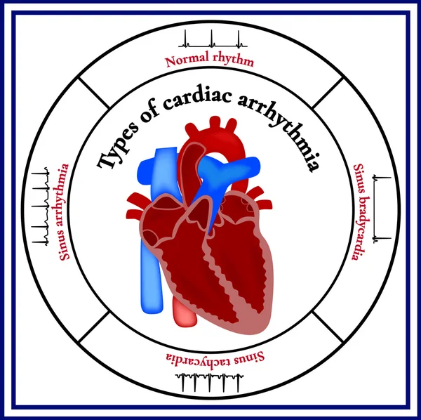
Stock image Atrial Tachycardia

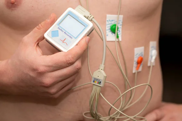
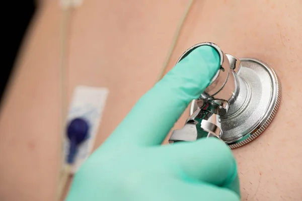
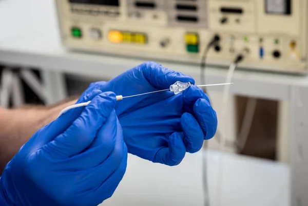
Cardiologist Use Tubes And Ultrasound For Radiofrequency Catheter Ablation.
Image, 6.54MB, 4801 × 3124 jpg

When The Rhythm Of The Atria Originates In The Lower Part Of The Atria, The Whole Atria Are Excited From Inferior To Superior, Producing Negative P Waves In The Inferior Leads.
Image, 11.92MB, 10000 × 8280 jpg
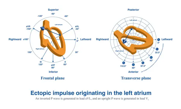
When Ectopic Impulses Originate In The Left Atrium, An Upright P Wave Will Be Generated In V1 And An Inverted P Wave In Lead AVL.
Image, 7.38MB, 10000 × 6060 jpg

Schemes Set Of Common Electrocardiogram (ECG) Abnormalities, Including Partial Blocks And Flutter
Vector, 9.68MB, 7750 × 4367 eps
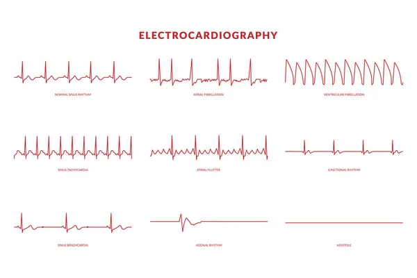
Electrocardiography Heartbeat Line Monitor. Vector EPS10 Illustration
Vector, 0.82MB, 6000 × 4000 eps


Treatment, Support With Medication And Heart Protection. Drugs - Vials And Syringe On Red Background Aimed At Heart, Which Lies Nearby. For Use In Cardiology And Treatment Of Cardiovascular System
Image, 4.65MB, 6016 × 4000 jpg

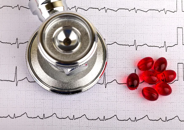
Diagnosis, Treatment And Prevention Of Diseases Of Heart And Cardiovascular System Concept Photo. Blue Stethoscope Is Surrounded By Tape Of ECG With Electrocardiogram Drawn On It. Diagnosis Of Disease
Image, 6.13MB, 6000 × 4000 jpg


Fast Heart And Rapid Heartbeat Or Pulse As A Cardiology Medical Concept As A Human Circulatory Organ Shaped As A Running Animal As A Cardiac Fatigue Idea With 3D Illustration Elements.
Image, 4.46MB, 4924 × 3680 jpg
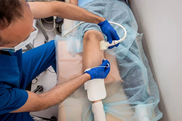
Cardiologist Use Tubes And Ultrasound For Radiofrequency Catheter Ablation.
Image, 6.73MB, 4740 × 3164 jpg
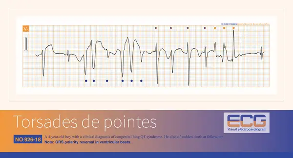
A 4-year-old Boy With A Clinical Diagnosis Of Long QT Syndrome. No Genetic Testing Was Done During Hospitalization. The Child Died Suddenly During Follow-up.
Image, 7.66MB, 10000 × 5414 jpg


Electromechanical Separation Is A Kind Of Terminal ECG. The Patient's ECG Has Electrical Signals, The ECG Wave Is Widened With Morphological Abnormalities, And The Ventricle Has No Contraction.
Image, 21.68MB, 10000 × 10515 jpg


Antiarrhythmic Drug For Treatment Or Suppress Abnormal Rhythm Of Heart, For Prevention Or Prophylaxis. Packing Of Pills With Inscription "Antiarrhythmic Medication"
Image, 11.18MB, 6000 × 4000 jpg

Cardiologist Use Tubes And Ultrasound For Radiofrequency Catheter Ablation.
Image, 6.45MB, 4505 × 3328 jpg
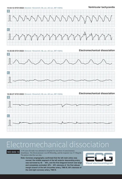
A Patient With Acute Extensive Anterior Myocardial Infarction Developed Ventricular Tachycardia During Hospitalization And Quickly Experienced Cardiac Arrest.
Image, 31.98MB, 10000 × 14632 jpg


Sotalol Drug Molecule. Used To Treat And Prevent Abnormal Heart Rhythms. Molecular Model. 3D Rendering. Illustration
Image, 5.41MB, 8700 × 4260 jpg

Sotalol Drug Molecule. Used To Treat And Prevent Abnormal Heart Rhythms. Skeletal Chemical Formula. Paper Packaging For Drugs. Vector Illustration
Vector, 0.32MB, 6239 × 4007 eps


Cardiologist Use Tubes And Ultrasound For Radiofrequency Catheter Ablation.
Image, 6.78MB, 4740 × 3164 jpg
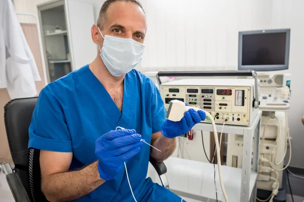
Cardiologist Use Tubes And Ultrasound For Radiofrequency Catheter Ablation.
Image, 5.86MB, 4740 × 3164 jpg

The Abnormal Fast Human Heart Increased Heart Rhythm Rate At Which It Was In Cardiac Arrest, Anxiety, Or Panic Attacks, 3D Render
Image, 0.48MB, 3840 × 2160 jpg


Sotalol Drug Molecule. Used To Treat And Prevent Abnormal Heart Rhythms. Structural Chemical Formula, Molecule Model. Vector
Vector, 0.38MB, 5661 × 4416 eps

Page 1 >> Next