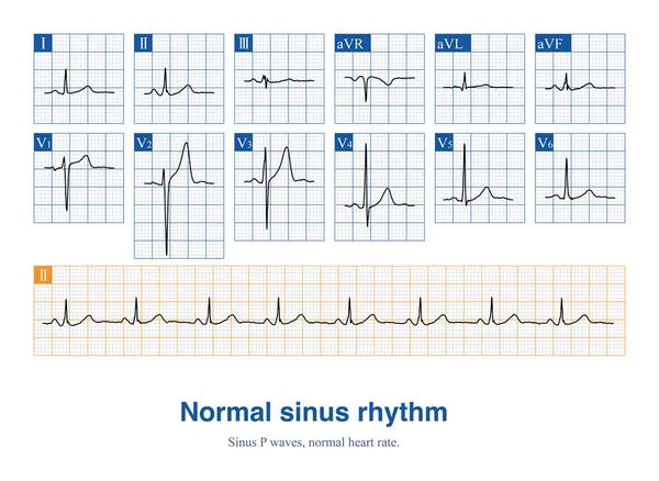
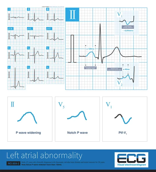
Stock image When the rhythm of the atria originates in the lower part of the atria, the whole atria are excited from inferior to superior, producing negative P waves in the inferior leads.

Published: Apr.23, 2024 12:49:32
Author: asia11m
Views: 1
Downloads: 1
File type: image / jpg
File size: 11.92 MB
Orginal size: 10000 x 8280 px
Available sizes:
Level: beginner