Home
Images
Vectors
Videos
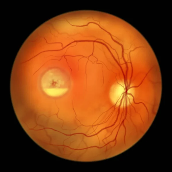
Stock image Best disease. Best vitelliform macular dystrophy, Pseudohypopyon stage, layering of lipofuscin, scientific illustration, ophthalmoscope view
Published:
Mar.16, 2023 08:26:23
Author:
katerynakon
Views:
10
Downloads:
1
File type:
image
/
jpg
File size:
2.89 MB
Orginal size:
5000
x
5000
px
Available sizes:
S
(500 x 500)
M
(1000 x 1000)
L
(2000 x 2000)
XL
(5000 x 5000)
EL0
(5000 x 5000)
Level:
silver
Download Image
Image topics:
Illustration
Best
Medical
Eye
Patient
Stage
Vessel
Pigment
Vision
Organ
Disk
Optic
Genetic
Investigation
Ophthalmologist
Disorder
Ocular
Ophthalmology
Retina
Ophthalmoscopy
Retinal
Hereditary
Fovea
Fundus
Macula
Macular
Hand Drawn
Hand Drawing
Epithelium
Dystrophy
Retinoscopy
Fundoscopy
Optic Disk
Retinal Dystrophy
Optic Cup
Bvmd
Bestrophinopathy
Vitelliform
Best 1 Gene
Bestrophin
Best Disease
Pseudohypopyon
Layering Of Lipofuscin
Similar stock images
Best Disease. Best Vitelliform Macular Dystrophy, Pseudohypopyon Stage, Layering Of Lipofuscin, Scientific Illustration, Ophthalmoscope View
5000 × 5000
Best Disease. Best Vitelliform Macular Dystrophy, Vitelleruptive Stage, Scrambled Egg Appearance, Scientific Illustration, Ophthalmoscope View
5000 × 5000
Non-proliferative Diabetic Retinopathy, Illustration Showing IRMAs (intraretinal Microvascular Abnormalities) As Small Vessels With Abnormal Branching Or Dilatation In Ischaemic Areas
5000 × 5000
Normal Eye Retina, Ophthalmoscope View, Scientific Illustration Showing Optic Disk, Blood Vessels, Macula And Fovea
5000 × 5000
Diabetic Macular Edema (DME). Diabetic Retinopathy, Illustration Showing Macula Edema, Abnormal Finding On Fundoscopic Examination Of The Eye Retina In Diabetes Mellitus
5000 × 5000
Non-proliferative Diabetic Retinopathy, Illustration Showing IRMAs (intraretinal Microvascular Abnormalities) As Small Vessels With Abnormal Branching In Ischaemic Areas, And Venous Beading
5000 × 5000
Non-proliferative Diabetic Retinopathy, Illustration Showing Small Retinal Haemorrhages, Ophthalmoscope View
5000 × 5000
Non-proliferative Diabetic Retinopathy, Illustration Showing Hard Exudates (irregularly Shaped Yellow Spots) And Microaneurysms (red Dots) On Funduscopic Examination Of The Eye Retina
5000 × 5000