Home
Images
Vectors
Videos
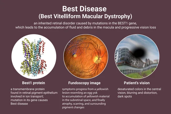
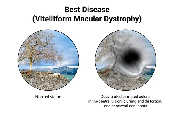
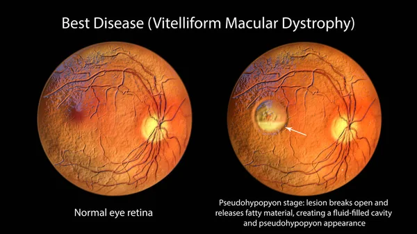
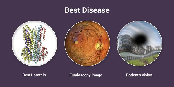
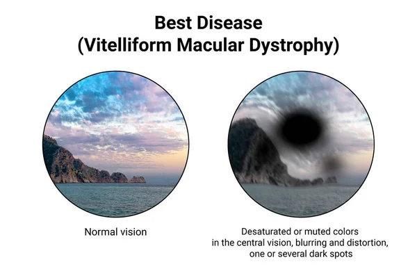
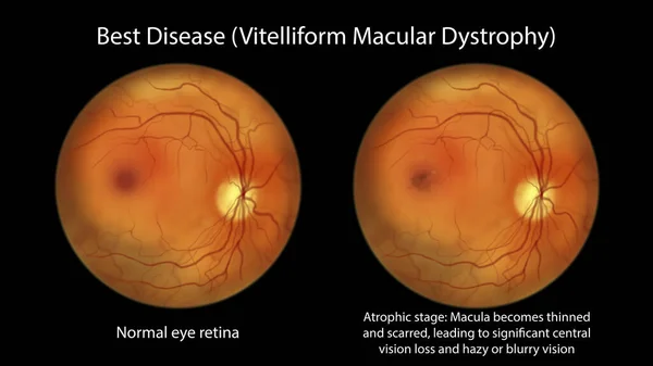
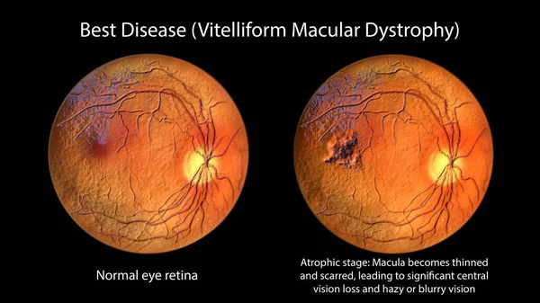
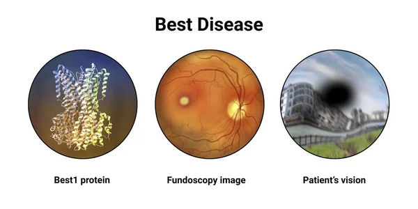
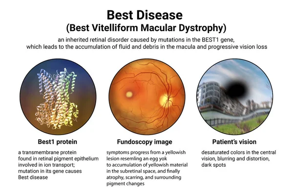
Stock image Best disease. Best vitelliform macular dystrophy, 3D illustration showing Best1 protein, classic fundoscopic egg-yolk lesion on retina, and distorted vision with black spot in a patient
Published:
Apr.04, 2023 11:54:01
Author:
katerynakon
Views:
65
Downloads:
2
File type:
image
/
jpg
File size:
7.56 MB
Orginal size:
9000
x
6000
px
Available sizes:
S
(500 x 333)
M
(1000 x 667)
L
(2000 x 1333)
XL
(9000 x 6000)
EL0
(9000 x 6000)
Level:
silver
Download Image
Image topics:
Illustration
Best
Medical
Eye
Patient
Stage
Vessel
Pigment
Vision
Organ
Disk
Optic
Genetic
Poster
Educational
Investigation
Ophthalmologist
Disorder
Ocular
Ophthalmology
Retina
Ophthalmoscopy
Retinal
Lesion
Hereditary
Fovea
Fundus
Macula
Macular
Egg Yolk
Hand Drawn
Hand Drawing
Epithelium
Dystrophy
Retinoscopy
Fundoscopy
Optic Disk
Retinal Dystrophy
Distorted Vision
Optic Cup
Bvmd
Bestrophinopathy
Vitelliform
Best 1 Gene
Bestrophin
Best Disease
Layering Of Lipofuscin
Best 1 Protein
Similar stock images
Best Disease. Best Vitelliform Macular Dystrophy, 3D Illustration Showing Best1 Protein, Classic Fundoscopic Egg-yolk Lesion On Retina, And Distorted Vision With Black Spot In A Patient
9000 × 6000
The Difference Between The Vision Of A Normal Eye And An Eye Affected By Best Disease, Illustration Showing Desatured Colors In The Central Vision, Blurring, Distortion, Black Spot
9000 × 6000
Best Disease, 3D Illustration Showing Normal Eye Retina And Best Vitelliform Macular Dystrophy, Pseudohypopyon Stage With Layering Of Lipofuscin, Ophthalmoscope View
11738 × 6603
Best Disease. Best Vitelliform Macular Dystrophy, 3D Illustration Showing Best1 Protein, Classic Fundoscopic Egg-yolk Lesion On Retina, And Distorted Vision With Black Spot In A Patient
9000 × 4500
The Difference Between The Vision Of A Normal Eye And An Eye Affected By Best Disease, Illustration Showing Desatured Colors In The Central Vision, Blurring, Distortion, Black Spot
9000 × 6000
Best Disease, An Illustration Showing Normal Eye Retina And Best Vitelliform Macular Dystrophy In Atrophic Stage With Retinal Atrophy And Scar Formation, Ophthalmoscope View
11738 × 6603
Best Disease, 3D Illustration Showing Normal Eye Retina And Best Vitelliform Macular Dystrophy In Atrophic Stage With Retinal Atrophy And Scar Formation, Ophthalmoscope View
11738 × 6603
Best Disease. Best Vitelliform Macular Dystrophy, 3D Illustration Showing Best1 Protein, Classic Fundoscopic Egg-yolk Lesion On Retina, And Distorted Vision With Black Spot In A Patient
9000 × 4500