
Stock image Cardiac Conduction System
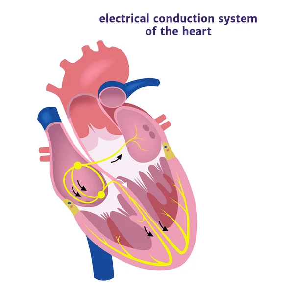
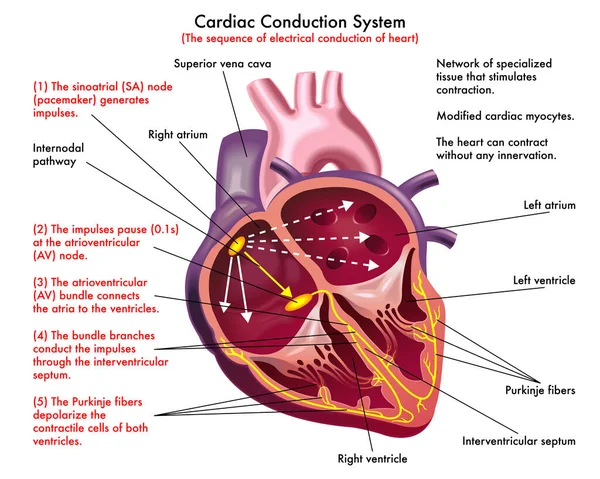
Diagram Of Cardiac Conduction System ( The Sequence Of Electrical Conduction Of Heart) With Annotations.
Vector, 3.47MB, 8858 × 7087 eps

In Complete Left Bundle Branch Block, The Conduction Of The LBB Can Be Completely Interrupted Or Can Still Be Conducted, But It Is Delayed By At Least 45ms Than The RBB.
Image, 10.8MB, 10000 × 5497 jpg

During Left Posterior Fascicular Block, The ECG Showed Right Axis Deviation. The QRS Wave In Leads I And AVL Was RS Wave, And The Duration Of QRS Wave Was Less Than 120 Ms.
Image, 30.53MB, 10000 × 11472 jpg

Doctor In Office At Desk With Laptop During Patient Admission Scans Paper Tape With Printed Result Of ECG. Concept Photo Of Diagnosis Of Cardiac Heart Disease, Arrhythmias, Heart Rhythm Disturbances
Image, 9.06MB, 6000 × 4000 jpg
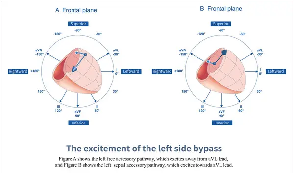
When The Left Free Wall And Septal Accessory Pathway Are Excited, Preexcitation Waves With Different Polarities Are Generated In Leads And AVL.
Image, 6.98MB, 14851 × 8810 jpg

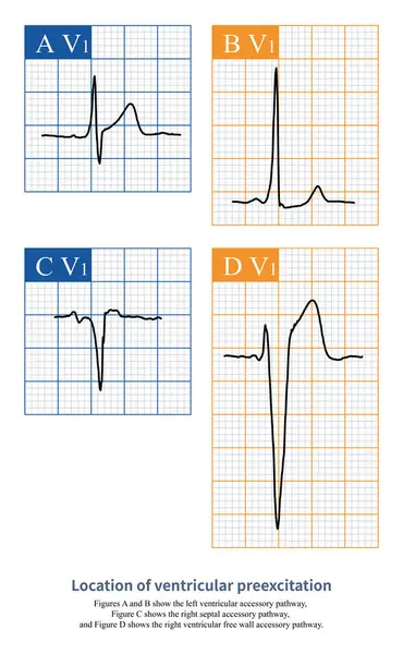
On The Electrocardiogram, Observing The Morphology Of QRS Waves In Lead V1 Can Distinguish Whether Ventricular Pre Excitation Is Located In The Left Ventricle Or The Right Ventricle.
Image, 4.78MB, 10000 × 11226 jpg

When The Ventricular Preexcitation Wave Leaves The Baseline And Then Falls Back To The Baseline, It Is Interpreted As An Isoelectric Line Preexcitation Wave.
Image, 9.3MB, 10000 × 11275 jpg

When The Left Anterior Wall And Posterior Wall Accessory Pathway Are Excited, Preexcitation Waves With Different Polarities Are Generated In The Inferior Wall Leads Of , And AVF.
Image, 3.66MB, 10000 × 5932 jpg

Ventricular Preexcitation Is The Pre Excitation Of A Portion Of The Ventricular Muscle By The Accessory Pathway, Forming A Rough And Dull, And Fuzzy Wave That Can Be Positive, Negative, Or Biphasic.
Image, 7.55MB, 10000 × 11891 jpg

Ventricular Preexcitation Is A Fusion Wave Formed By The Accessory Pathway And Normal Atrioventricular Conduction System Exciting A Part Of Ventricle Respectively.
Image, 4.25MB, 10000 × 10000 jpg

Abnormal ECG Refers To Changes In Depolarization Waves And Or Repolarization Waves, Most Of Which Are Pathologic And Few Are Physiological.
Image, 7.91MB, 10000 × 5625 jpg

When Ventricular Preexcitation Components Occupy Different Proportions Of QRS Waves, The Measured PJ Intervals Are Different.
Image, 4.21MB, 10000 × 4226 jpg

On The Electrocardiogram, Observing The Morphology Of QRS Waves In Lead V1 Can Distinguish Whether Ventricular Pre Excitation Is Located In The Left Ventricle Or The Right Ventricle.
Image, 18.67MB, 10000 × 14900 jpg

The Polarity Of Ventricular Preexcitation Waves Can Be Positive, As Shown In Figures A And B, Or Negative, As Shown In Figures C And D.
Image, 2.82MB, 10000 × 11593 jpg

When There Is A Left Ventricular Free Wall Bypass, The Polarity Of The Ventricular Preexcitation Is Positive In Lead V1 And Negative In Lead AVL On The Electrocardiogram.
Image, 7.36MB, 10000 × 6759 jpg
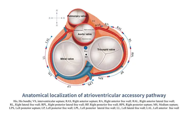
Surrounding The Atrioventricular Ring, Except For The Anterior Septum Of The Left Ventricle, There Is No Distribution Of Accessory Pathways, And Accessory Pathways Can Exist In Other Parts.
Image, 5.26MB, 10000 × 6257 jpg

Sometimes, There May Be Slight Non-specific Changes And Normal Variations In The Electrocardiogram, Which Are Often Due To Physiological Reasons And Have No Clinical Therapeutic Significance.
Image, 13.15MB, 10000 × 11438 jpg

Abnormal ECG Refers To Changes In Depolarization Waves And Or Repolarization Waves, Most Of Which Are Pathologic And Few Are Physiological.
Image, 13.09MB, 10000 × 11438 jpg

On The Electrocardiogram, Observing The Morphology Of QRS Waves In Lead V1 Can Distinguish Whether Ventricular Pre Excitation Is Located In The Left Ventricle Or The Right Ventricle.
Image, 2.99MB, 10000 × 6863 jpg

A 2:1 Left Bundle Branch Block Is Considered When Complete Left Bundle Branch Block Alternates With Normal QRS Complexes And The PR Interval Is Fixed.
Image, 5.72MB, 10000 × 3162 jpg

At The Beginning Of Heart Development, The Atria And Ventricles Form A Whole, Known As The Primitive Heart Tube.
Image, 4.96MB, 10000 × 10005 jpg
On The Electrocardiogram, Observing The Morphology Of QRS Waves In Lead V1 Can Distinguish Whether Ventricular Pre Excitation Is Located In The Left Ventricle Or The Right Ventricle.
Image, 7.46MB, 8000 × 12972 jpg

The Conduction In Ventricle Is Mainly Divided Into Right Bundle Branch And Left Bundle Branch. The Left Bundle Branch Includes Left Anterior Fascicle And Left Posterior Fascicle.
Image, 4.97MB, 10000 × 10000 jpg
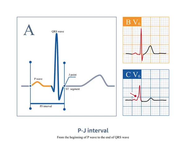
On The Electrocardiogram, The PJ Interval Is Used To Distinguish Between Ventricular Heartbeats And Ventricular Pre Excitation. The Normal Value Of PJ Interval Is Less Than 270ms.
Image, 8.38MB, 10000 × 8409 jpg

The AVN And His Bundle Form The Conduction Axis At The Atrioventricular Junction Region. The His Bundle Is Divided Into Three Parts: Non Penetrating Part, Penetrating Part, And Bifurcation Section.
Image, 4.89MB, 9000 × 12889 jpg


Cardiovascular System Heart Isolated Icon Internal Organ Vector Anatomy Physiology Heartbeat And Pulse Or Rate Blood Pressure And Flow Anatomical Structure Cardiology Medicine And Healthcare Biology.
Vector, 1.22MB, 5556 × 6396 eps


Human Heart Anatomy. Organs Symbol. Vector Illustration In Cartoon Style Isolated On White Background
Vector, 5.58MB, 2428 × 2368 eps


Page 1 >> Next