photo free
Home Images Vectors Videos

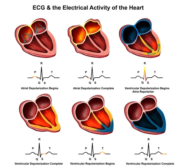
Stock image Abnormal ECG refers to changes in depolarization waves and or repolarization waves, most of which are pathologic and few are physiological.

Stock image Abnormal ECG refers to changes in depolarization waves and or repolarization waves, most of which are pathologic and few are physiological.
Published: Apr.18, 2024 16:32:40
Author: asia11m
Views: 10
Downloads: 1
File type: image / jpg
File size: 7.91 MB
Orginal size: 10000 x 5625 px
Available sizes:
S
(500 x 281)
M
(1000 x 563)
L
(2000 x 1125)
XL
(10000 x 5625)
EL0
(10000 x 5625)
Level: beginner
Download Image
Image topics:

Similar stock images

stock image Abnormal ECG refers to changes in depolarization waves and or repolarization waves, most of which are pathologic and few are physiological.

Abnormal ECG Refers To Changes In Depolarization Waves And Or Repolarization Waves, Most Of Which Are Pathologic And Few Are Physiological.

10000 × 11438
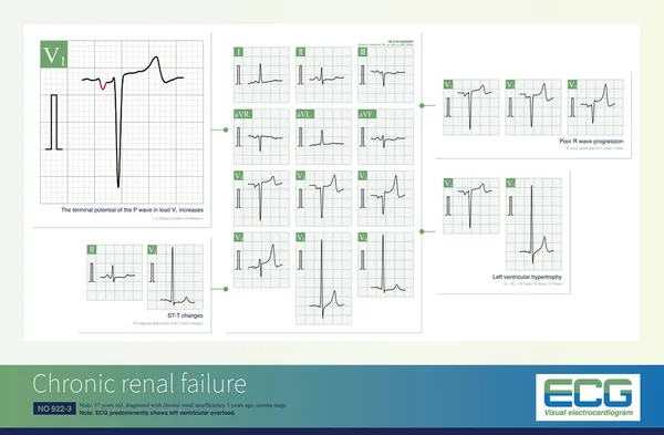
stock image Common ECG changes in chronic renal insufficiency include left ventricular hypertrophy, left atrial abnormality, poor R wave progression and ST-T changes.

Common ECG Changes In Chronic Renal Insufficiency Include Left Ventricular Hypertrophy, Left Atrial Abnormality, Poor R Wave Progression And ST-T Changes.

10000 × 6551
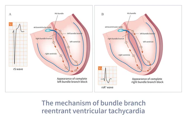
stock image The reentry of bundle branch reentrant ventricular tachycardia occurs between the left bundle branch and the right bundle branch. This is a malignant ventricular tachycardia.

The Reentry Of Bundle Branch Reentrant Ventricular Tachycardia Occurs Between The Left Bundle Branch And The Right Bundle Branch. This Is A Malignant Ventricular Tachycardia.

10000 × 6444
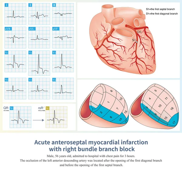
stock image When acute anteroseptal myocardial infarction combined with right bundle branch block, the QRS wave in lead V1 shows typical QR wave, and the initial r wave is lost.

When Acute Anteroseptal Myocardial Infarction Combined With Right Bundle Branch Block, The QRS Wave In Lead V1 Shows Typical QR Wave, And The Initial R Wave Is Lost.

10000 × 9474
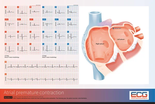
stock image Female, 57 years old, diagnosed with mitral stenosis 6 years ago. This electrocardiogram had an atrial premature contraction, indicating its origin in the right superior pulmonary vein.

Female, 57 Years Old, Diagnosed With Mitral Stenosis 6 Years Ago. This Electrocardiogram Had An Atrial Premature Contraction, Indicating Its Origin In The Right Superior Pulmonary Vein.

10000 × 6733
stock image Coronary artery spasm causes transmural myocardial ischemia, and ST segment elevation in ECG has localization characteristics. Criminal vessels can be derived from ST segment elevation leads in ECG.

Coronary Artery Spasm Causes Transmural Myocardial Ischemia, And ST Segment Elevation In ECG Has Localization Characteristics. Criminal Vessels Can Be Derived From ST Segment Elevation Leads In ECG.

10000 × 7684
stock image  Medical illustration of  ECG explained

Medical Illustration Of ECG Explained

8031 × 7471