
Stock image Caudata
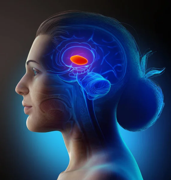
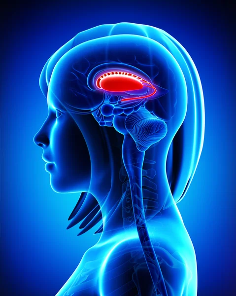
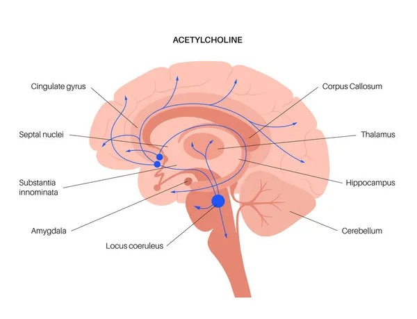
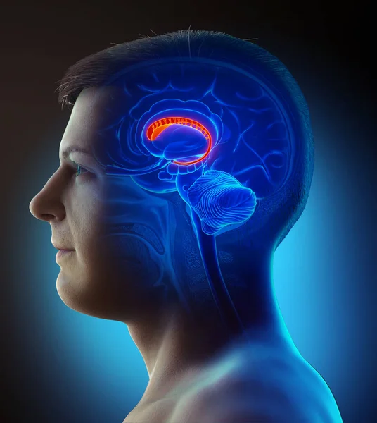
3d Rendered Medically Accurate Illustration Of A Female Brain Anatomy- The Thalamus
Image, 6.81MB, 4000 × 4200 jpg
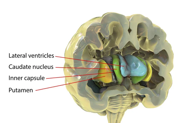
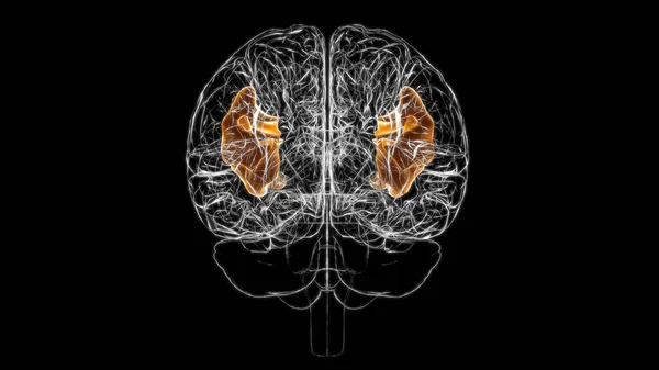
Human Brain Anatomy, Basal Ganglia. 3D Illustration Showing Caudate Nucleus (green), Putamen (yellow), And Lateral Ventricles (blue)
Image, 6.25MB, 6252 × 4168 jpg
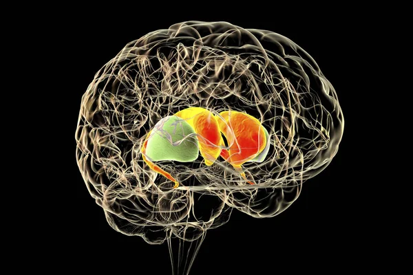
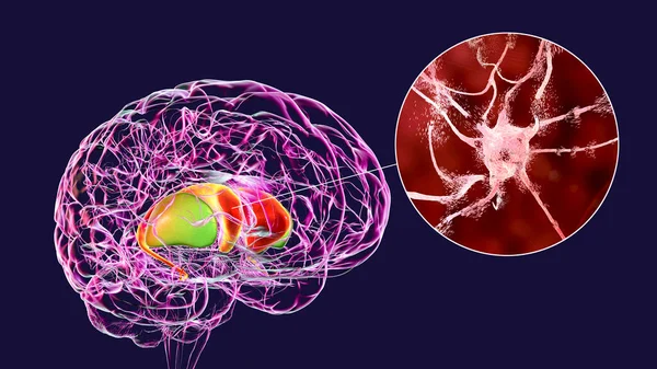
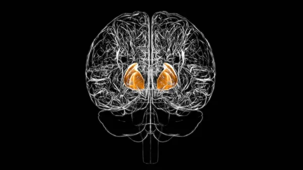
Dorsal Striatum In The Human Brain, 3D Illustration. It Is A Nucleus In The Basal Ganglia, Consists Of The Caudate Nucleus (red) And The Putamen (green), Is A Component Of The Motor And Reward Systems
Image, 6.21MB, 6000 × 4000 jpg
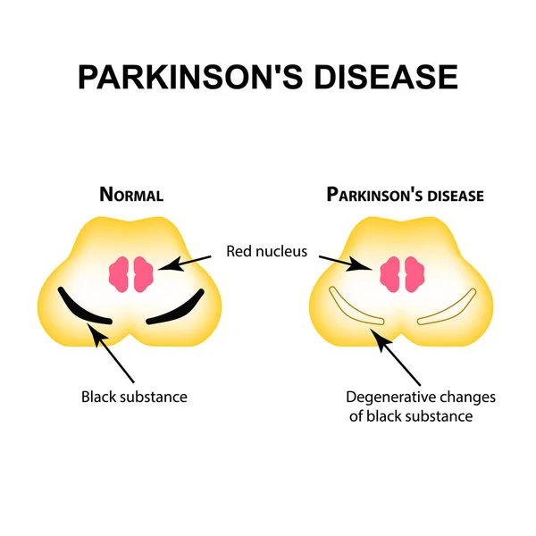
Parkinson's Disease. Degenerative Changes In The Brain Are A Black Substance. Vector Illustration On Isolated Background.
Vector, 0.95MB, 5000 × 5000 eps

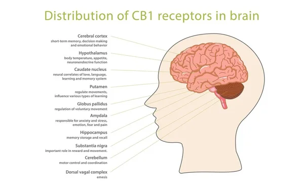
Cortex As A Complex Subject, Related To Important Topics. Pictured As A Puzzle And A Word Cloud Made Of Most Important Ideas And Phrases Related To Cortex.
Image, 2.74MB, 7680 × 4320 jpg


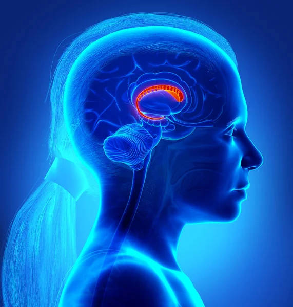
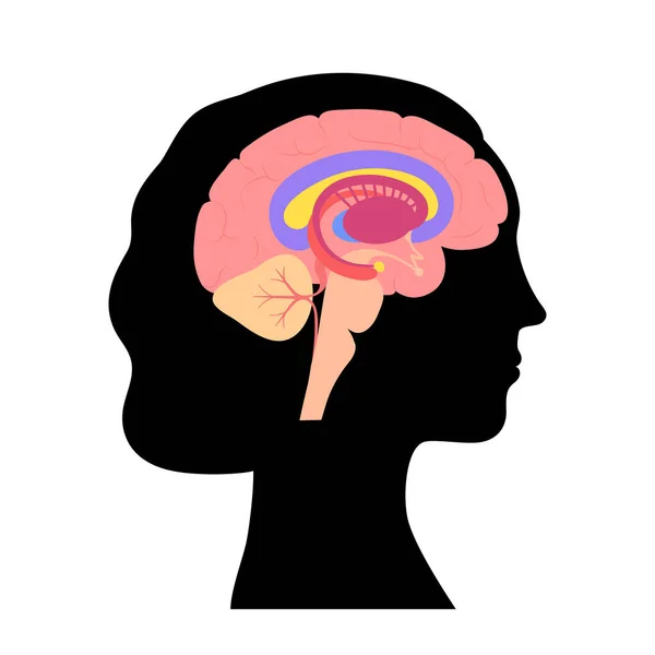
3d Rendered Medically Accurate Illustration Of A Young Girl Brains Anatomy-the Caudate Nucleus
Image, 7.67MB, 4000 × 4200 jpg
Dorsal Striatum Highlighted In Human Brain And Close-up View Of Degrading Neurons Of Dorsal Striatum Seen In Huntington's Disease, 3D Illustration
Image, 16.37MB, 8672 × 4878 jpg

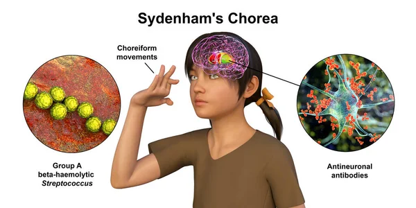
Sydenham's Chorea, An Autoimmune Disease That Results From Streptococcus Infection, Formation Of Anti-neuronal Antibodies Damaging Brain Basal Ganglia That Cause Involuntary Movements, 3D Illustration
Image, 12.54MB, 8488 × 4244 jpg

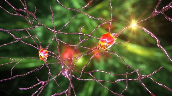
Neurons Of Dorsal Striatum, 3D Illustration. The Dorsal Striatum Is A Nucleus In The Basal Ganglia, Degrading Of Its Neurons Plays A Crucial Role In The Development Of Huntington's Disease
Image, 10.79MB, 7200 × 4050 jpg
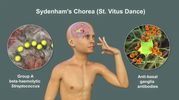
Sydenham's Chorea, An Autoimmune Disease That Results From Streptococcus Infection, Formation Of Anti-neuronal Antibodies Damaging Brain Basal Ganglia That Cause Involuntary Movements, 3D Illustration
Image, 16.36MB, 8737 × 4914 jpg
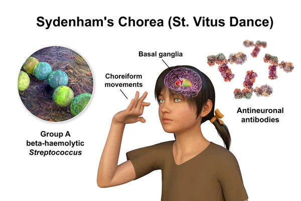
Sydenham's Chorea, An Autoimmune Disease That Results From Streptococcus Infection, Formation Of Anti-neuronal Antibodies Damaging Brain Basal Ganglia That Cause Involuntary Movements, 3D Illustration
Image, 10.04MB, 7883 × 5255 jpg


Dorsal Striatum Highlighted In Child's Brain And Close-up View Of Its Neurons, 3D Illustration. It Is A Nucleus In The Basal Ganglia, A Component Of The Motor And Reward Systems
Image, 31.18MB, 9339 × 6226 jpg
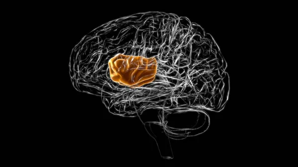
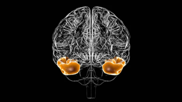
Brain Inferior Temporal Gyrus Anatomy For Medical Concept 3D Illustration
Image, 1.79MB, 3840 × 2160 jpg

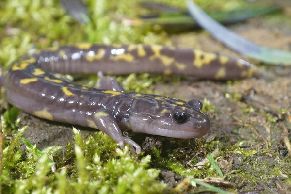
Closeup On The Critically Endangered Gorgan Mountain Salamander, Paradactylodon Gorganensis On Green Moss
Image, 5.26MB, 3000 × 2000 jpg

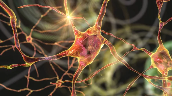
Neurons Of Dorsal Striatum, 3D Illustration. The Dorsal Striatum Is A Nucleus In The Basal Ganglia, Degrading Of Its Neurons Plays A Crucial Role In The Development Of Huntington's Disease
Image, 12.53MB, 7200 × 4050 jpg
Brain Inferior Frontal Gyrus Anatomy For Medical Concept 3D Illustration
Image, 1.78MB, 3840 × 2160 jpg
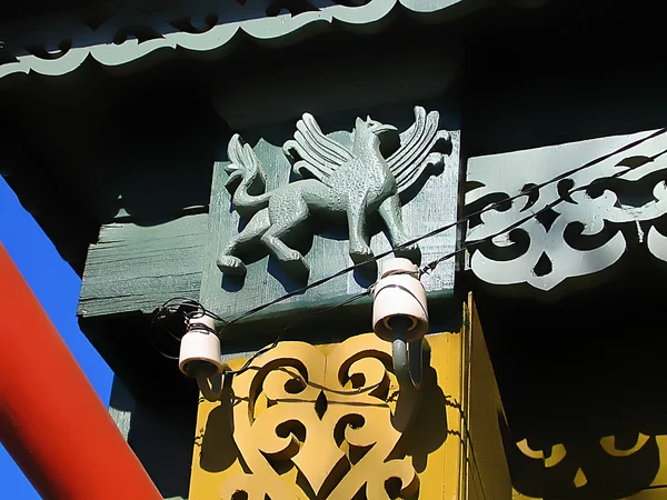
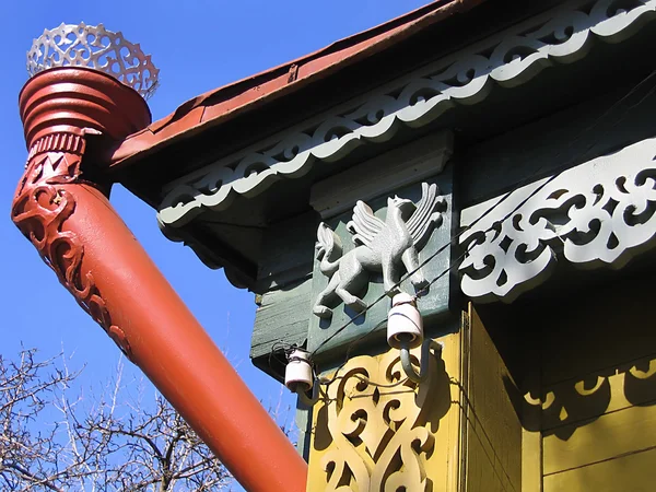
Fragment Of A Old Russian House With Decorative Carvings And The Figure Of A Griffin. Shot Taken In Kolomna - Provincial Russian Town
Image, 3.16MB, 2816 × 2112 jpg


Parkinson Disease - Diagnosis Written On A White Piece Of Paper. Syringe And Vaccine With Drugs.
Image, 7.35MB, 5010 × 3398 jpg

3d Rendered Medically Accurate Illustration Of A Male Brain Anatomy-the Lateral Globus Pallidus
Image, 3.86MB, 3669 × 4200 jpg
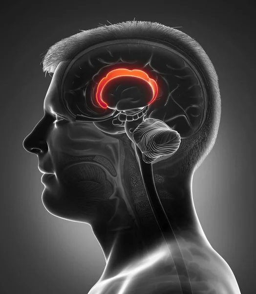
3d Rendered Medically Accurate Illustration Of A Male Brains Anatomy-the Caudate Nucleus
Image, 5.44MB, 3758 × 4200 jpg


Dorsal Striatum Highlighted In Child's Brain And Close-up View Of Its Neurons, 3D Illustration. It Is A Nucleus In The Basal Ganglia, A Component Of The Motor And Reward Systems
Image, 23.52MB, 9339 × 6226 jpg

Destruction Of Neurons Of The Caudate Nucleus, Conceptual 3D Illustration. Caudate Nucleus Belongs To The Brain Basal Ganglia, Its Neurons Are Damaged In Huntingon's Disease And Other Choreas
Image, 16.49MB, 7996 × 5331 jpg

Page 1 >> Next