photo free
Home Images Vectors Videos

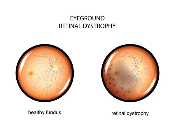
Stock image Retinal Dystrophy

stock image Blue female eye on surgery clinic exam closeup

Blue Female Eye On Surgery Clinic Exam Closeup

Image, 5.41MB, 5244 × 3496 jpg
stock image Best disease. Best vitelliform macular dystrophy, Vitelliform stage, classic egg-yolk lesion, scientific illustration, ophthalmoscope view

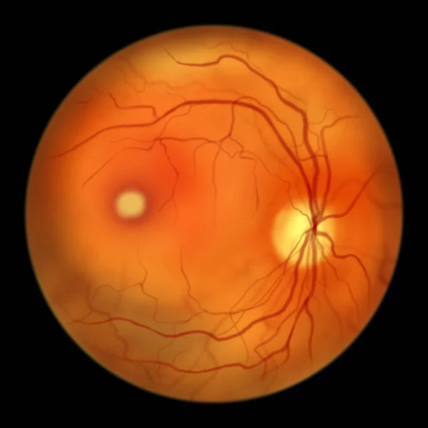
Best Disease. Best Vitelliform Macular Dystrophy, Vitelliform Stage, Classic Egg-yolk Lesion, Scientific Illustration, Ophthalmoscope View

Image, 2.87MB, 5000 × 5000 jpg
stock image Best disease. Best vitelliform macular dystrophy, 3D illustration showing Best1 protein, classic fundoscopic egg-yolk lesion on retina, and distorted vision with black spot in a patient

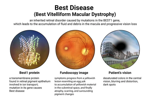
Best Disease. Best Vitelliform Macular Dystrophy, 3D Illustration Showing Best1 Protein, Classic Fundoscopic Egg-yolk Lesion On Retina, And Distorted Vision With Black Spot In A Patient

Image, 7.56MB, 9000 × 6000 jpg
stock image Best disease. Best vitelliform macular dystrophy, Pseudohypopyon stage, layering of lipofuscin, scientific illustration, ophthalmoscope view

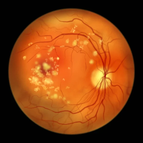
Best Disease. Best Vitelliform Macular Dystrophy, Pseudohypopyon Stage, Layering Of Lipofuscin, Scientific Illustration, Ophthalmoscope View

Image, 2.89MB, 5000 × 5000 jpg
stock image Best disease, illustration showing normal eye retina and Best vitelliform macular dystrophy, Vitelleruptive stage with scrambled egg appearance on fluorescein angiography

Best Disease, Illustration Showing Normal Eye Retina And Best Vitelliform Macular Dystrophy, Vitelleruptive Stage With Scrambled Egg Appearance On Fluorescein Angiography

Image, 8.14MB, 11738 × 6603 jpg
stock image Autosomal recessive bestrophinopathy, ophthalmoscope view, scientific illustration showing accumulation of lipofuscin deposits around and beyond the macula leading to progressive damage to the retina

Autosomal Recessive Bestrophinopathy, Ophthalmoscope View, Scientific Illustration Showing Accumulation Of Lipofuscin Deposits Around And Beyond The Macula Leading To Progressive Damage To The Retina

Image, 3MB, 5000 × 5000 jpg
stock image The difference between the vision of a normal eye and an eye affected by Best disease, illustration showing desatured colors in the central vision, blurring, distortion, black spot

The Difference Between The Vision Of A Normal Eye And An Eye Affected By Best Disease, Illustration Showing Desatured Colors In The Central Vision, Blurring, Distortion, Black Spot

Image, 12.33MB, 9000 × 6000 jpg
stock image Retinitis pigmentosa, a genetic eye disease. An illustration shows normal eye retina and attenuated blood vessels, pigmentary bone spicules and waxy appearance of the optic disk in the affected retina

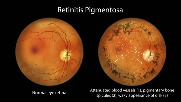
Retinitis Pigmentosa, A Genetic Eye Disease. An Illustration Shows Normal Eye Retina And Attenuated Blood Vessels, Pigmentary Bone Spicules And Waxy Appearance Of The Optic Disk In The Affected Retina

Image, 7.9MB, 11738 × 6603 jpg
stock vector fundus. retinal tears

Fundus. Retinal Tears

Vector, 6.78MB, 4800 × 3700 eps
stock vector Retinal detachment. Cross section of a normal human eyes and disorder of the eye in which the retina peels. Schematic diagram. Detailed vector illustration

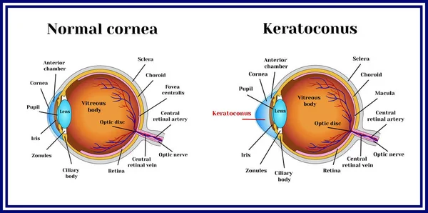
Retinal Detachment. Cross Section Of A Normal Human Eyes And Disorder Of The Eye In Which The Retina Peels. Schematic Diagram. Detailed Vector Illustration

Vector, 4.17MB, 6309 × 3000 eps
stock vector Hand drawn eyeball with retinal tear and retinal detachment. Disease of the organ of vision. Vector illustration.

Hand Drawn Eyeball With Retinal Tear And Retinal Detachment. Disease Of The Organ Of Vision. Vector Illustration.

Vector, 0.35MB, 5679 × 3096 eps
stock image Female eye on surgery clinic exam closeup

Female Eye On Surgery Clinic Exam Closeup

Image, 3.95MB, 6720 × 2240 jpg
stock image Female eye on surgery clinic exam closeup

Female Eye On Surgery Clinic Exam Closeup

Image, 7.22MB, 6720 × 4480 jpg
stock vector eye, fundus. retinitis

Eye, Fundus. Retinitis

Vector, 13.13MB, 4800 × 3700 eps
stock vector Keratoconus. Dystrophic disease of the cornea.

Keratoconus. Dystrophic Disease Of The Cornea.

Vector, 0.8MB, 7087 × 3528 eps
stock vector Keratoconus. Dystrophic disease of the cornea.

Keratoconus. Dystrophic Disease Of The Cornea.

Vector, 0.62MB, 5315 × 5315 eps
stock vector fundus. retinal vascular thrombosis

Fundus. Retinal Vascular Thrombosis

Vector, 12.95MB, 4801 × 3601 eps
stock vector eyeground. retinal dystrophy

Eyeground. Retinal Dystrophy

Vector, 6.14MB, 4801 × 3601 eps
stock image Ray of light falls on human eye

Ray Of Light Falls On Human Eye

Image, 13.28MB, 6720 × 4480 jpg
stock vector fundus. pigment retinopathy

Fundus. Pigment Retinopathy

Vector, 10.42MB, 4800 × 3700 eps
stock vector fundus. epiretinal membrane

Fundus. Epiretinal Membrane

Vector, 9.91MB, 4800 × 3700 eps
Page 1 >> Next