Home
Images
Vectors
Videos
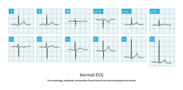
Stock image Because of the slow conduction of atrioventricular node, the PR interval of adult ECG should be greater than 120ms. This physiological phenomenon is called atrioventricular delay.
Published:
May.18, 2024 06:55:01
Author:
asia11m
Views:
0
Downloads:
0
File type:
image
/
jpg
File size:
8.27 MB
Orginal size:
10000
x
6886
px
Available sizes:
S
(500 x 344)
M
(1000 x 689)
L
(2000 x 1377)
XL
(10000 x 6886)
EL0
(10000 x 6886)
Level:
beginner
Download Image
Image topics:
Graphic
Human
Health
Medicine
Medical
Care
Wave
Cardiology
Heart
Hospital
Disease
Education
Death
Graph
Beat
Rhythm
Diagnosis
Heartbeat
Anatomy
Electrode
Cardiogram
Cardiac
Ekg
Electrocardiogram
Atrium
Ventricle
Arrhythmia
Electrocardiograph
Heart Disease
Action Potential
Conduction System
ECG Monitoring
Cardiac Anatomy
Sinoatrial Node
Atrioventricular Node
Cardiovascular Medicine
St Segment
Ecg Theory
Ecg Education
Qrs Wave
Purkinje Fiber
His Bundle
Bundle Branch
Refractory Period
Descending Conduction
Slow Conduction
Physiological Phenomenon
Atrioventricular Delay
Similar stock images
Acute Left Main Artery Occlusion Can Cause Both ST Segment Elevation And Non ST Segment Elevation Myocardial Infarction, Regardless Of Which Type, The Risk Of Death Is High.
10000 × 5399
Atrioventricular Separation Is A Clue To The Diagnosis Of Ventricular Tachycardia. The Grass Yellow Arrow Shows The Sinus P Wave, Which Is Obviously Not Related To The QRS Wave.
10000 × 3338
The Left Ventricular Wall Activation Time Is The Time Interval Measured From The Beginning Of The QRS Wave To The Peak Of The R Wave.
10000 × 10905
In Some Patients With Acute Anterior Myocardial Infarction, The Inferior Leads Of The Electrocardiogram Will Have ST Segment Depression, Which Is Called Corresponding ST Segment Depression.
10000 × 6954
When Acute Left Main Artery Occlusion Occurs, QRS Wave Of ECG Can Be Normal, Or Wide QRS Wave Can Occur With Intraventricular Block, And The Prognosis Of The Latter Is Worse.
10000 × 8109
The ECG Changes Of Left Atrium Abnormality Include Widening Of P Wave Duration, Bimodal P Wave And Increasing Of P Wave Terminal Potential In Lead V1 Lead P Wave.
10000 × 8526
In AMI, If The Amplitude Of ST Segment Elevation Is Less Than 1mm, It Is Called Slight ST Segment Elevation Myocardial Infarction, Which Is Easily Misdiagnosed As Early Repolarization
7000 × 9341
The Normal Rhythm Is Sinus Rhythm.The Morphology, Amplitude, And Duration Of Each Wave In The Electrocardiogram Are Normal.
10000 × 4964