Home
Images
Vectors
Videos
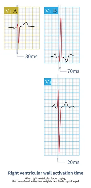
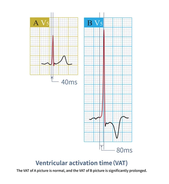
Stock image The left ventricular wall activation time is the time interval measured from the beginning of the QRS wave to the peak of the R wave.
Published:
May.12, 2023 13:01:04
Author:
asia11m
Views:
10
Downloads:
0
File type:
image
/
jpg
File size:
12.23 MB
Orginal size:
10000
x
10905
px
Available sizes:
S
(459 x 500)
M
(917 x 1000)
L
(1834 x 2000)
XL
(10000 x 10905)
EL0
(10000 x 10905)
Level:
beginner
Download Image
Image topics:
Blue
Graphic
Illustration
Rate
Human
Health
Healthy
Medicine
Medical
Cardiology
Heart
Hospital
Pulse
Body
Disease
Education
Science
Death
Graph
Beat
Rhythm
Diagnosis
Heartbeat
Cardiogram
Cardiac
Ekg
Electrocardiogram
Ecg
Electrocardiograph
Hypertrophy
Myocardium
Heart Disease
Chest Pain
Cardiac Arrest
Cardiomyopathy
Left Ventricle
Sudden Death
Dilated Cardiomyopathy
Myocardial Ischemia
ECG Monitoring
Left Ventricular Hypertrophy
P Wave
St Segment
Ecg Education
Qrs Wave
T Wave Inversion
Hypertrophic Cardiomyopathy
St Changing
Similar stock images
When Right Ventricular Hypertrophy, The Time Of Wall Activation In Right Chest Leads Is Prolonged.Figure A Shows The Normal Control, And Figure B Shows Right Ventricle Hypertrophy.
8000 × 16564
When The Left Ventricular Volume Load Increases, The ECG Not Only Shows Changes In Left Ventricular Hypertrophy, But Also Increases The Amplitude Of The T Wave.
10000 × 10000
Atrial Depolarization Forms A P Wave, Ventricular Depolarization Forms A QRS Wave, And Ventricular Repolarization Forms A T Wave, Which Constitutes A Common Waveform Of The Electrocardiogram.
10000 × 9917
In AMI, If The Amplitude Of ST Segment Elevation Is Less Than 1mm, It Is Called Slight ST Segment Elevation Myocardial Infarction, Which Is Easily Misdiagnosed As Early Repolarization
7000 × 9341
Generally, The Amplitude Of ST Segment Elevation Of AVR Lead 1mm Is Clinically Significant, And It Is Often Used For Criminal Vessel Locationand Prognosis Interpretation Of Acute Coronary Syndrome.
10000 × 9984
If Atrioventricular Dissociation And Ventricular Capture Are Found During Wide QRS Tachycardia, It Supports The Diagnosis Of Ventricular Tachycardia With High Specificity And Poor Sensitivity.
10000 × 3269
Sometimes, Because The QRS Axis Is In The Upper Left Quadrant, The High-amplitude R Wave Of Left Ventricular Hypertrophy Occurs In The Limb Leads, And Left Chest Leads Is Normal.
10000 × 10111