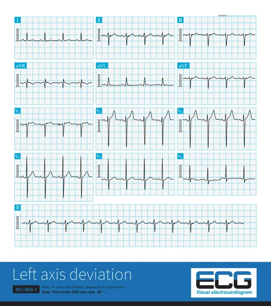
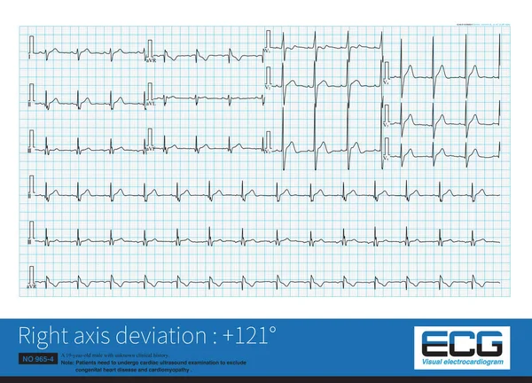
Stock image In complete left bundle branch block, the QRS wave in lead V1 is usually in rS and QS morphologies, with QRS duration 120ms, which is common in organic heart disease.

Published: Oct.06, 2022 07:24:05
Author: asia11m
Views: 18
Downloads: 0
File type: image / jpg
File size: 20.36 MB
Orginal size: 10000 x 7070 px
Available sizes:
Level: beginner