Home
Images
Vectors
Videos
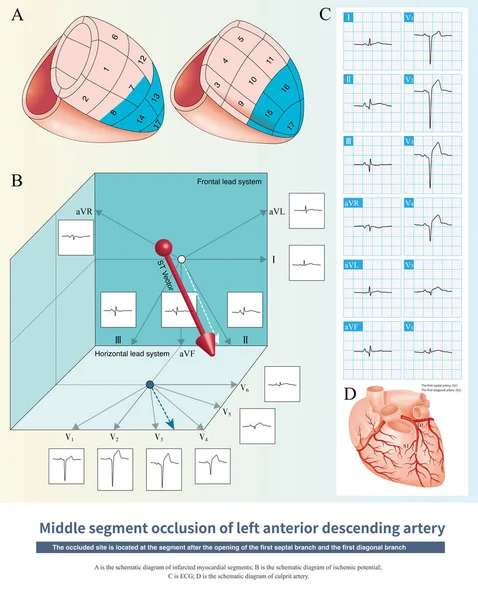
Stock image When the middle - distal segments of the LAD were occluded, the ischemic potential was toward the anterior, inferior and left, and the infarct spread to the anterior lower part of the left ventricle.
Published:
Mar.15, 2023 15:18:40
Author:
asia11m
Views:
34
Downloads:
0
File type:
image
/
jpg
File size:
17.25 MB
Orginal size:
9000
x
11279
px
Available sizes:
S
(399 x 500)
M
(798 x 1000)
L
(1596 x 2000)
XL
(9000 x 11279)
EL0
(9000 x 11279)
Level:
beginner
Download Image
Image topics:
Graphic
Illustration
Design
Human
Healthy
Medicine
Medical
Cardiology
Heart
Body
Disease
Education
Science
Death
Vessel
Organ
Diagnosis
Anatomy
Cardiac
Ekg
Electrocardiogram
Ecg
Anatomical
Atherosclerosis
Arteriosclerosis
Heart Disease
Chest Pain
Cardiac Arrest
Coronary Artery
Left Ventricle
Sudden Death
Ischemic Heart Disease
Myocardial Ischemia
Conduction System
Acute Coronary Syndrome
ECG Monitoring
Conduction Disorder
Bundle Branch Block
Collateral Circulation
Coronary Spasm
Ecg Education
Left Anterior Descending Artery
Ischemic Chest Pain
Left Main Trunk
Right Bundle Branch
Bundle Branch
Luminal Stenosis
Diagonal Artery
Anteroseptal Infarction
Similar stock images
When The Occlusion Of The LAD Affects The First Septal Branch And The First Diagonal Branch Is Not Affected, The Ischemic Potential Mainly Faces Anterior, Right And Inferior.
10000 × 12578
When The Middle-distal Segments Of The LAD Are Occluded, The ECG Of Extensive Anterior Myocardial Infarction Will Also Be Generated, But The Infarction Range Is Less Than That Of High Occlusion.
10000 × 8439
When The Proximal To Middle Segment Of The Right Coronary Artery Is Occluded, The Ischemic Potential Faces Inferior And Posterior, And Can Be Left Or Right, As Shown In The Diagram To The Right.
10000 × 11845
When Both High Lateral And Inferior Myocardial Infarction Occur Simultaneously, Electrocardiographic ST-T May Be Normal Due To The Combination Of Ischemic Elevation And Corresponding Depression.
10000 × 8197
In The Case Of Acute Anterior Myocardial Infarction, The Amplitude Of ST Segment Offset Can Be Used To Deduce That The Occlusive Site Is Located In The Proximal Segment Of The LAD.
10000 × 8502
After The First Diagonal Branch And Before The First Septal Branch Of The LAD Were Occluded, The Infarction Affected The Anterior Septum, And The High Lateral Wall Was Not Affected.
10000 × 8484
When Extensive ST Segment Depression Occurs, The Most Important Differential Diagnosis Of ECG Is To Deduce Left Main Artery Occlusion Or Single Coronary Artery Occlusion.
10000 × 6825
After The First Diagonal Branch And Before The First Septal Branch Of The LAD Were Occluded, The Infarction Affected The Anteroseptum, And The High Lateral Wall Was Not Affected.
10000 × 8459