Home
Images
Vectors
Videos
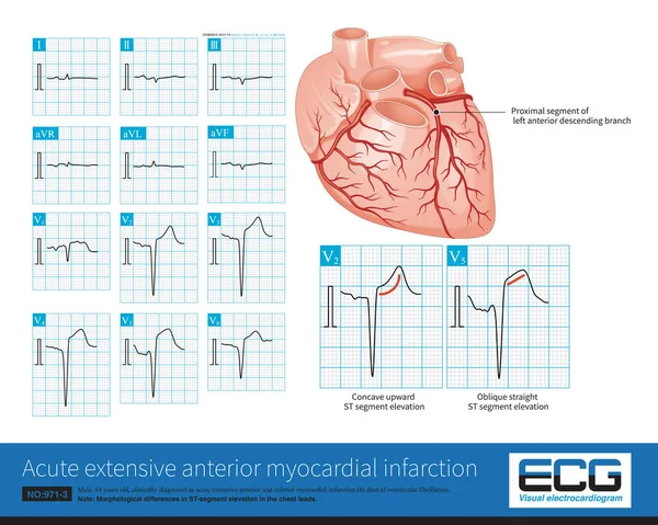
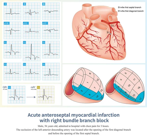
Stock image When acute anteroseptal myocardial infarction combined with right bundle branch block, the QRS wave in lead V1 shows typical QR wave, and the initial r wave is lost.
Published:
Mar.15, 2023 09:05:27
Author:
asia11m
Views:
365
Downloads:
0
File type:
image
/
jpg
File size:
18.51 MB
Orginal size:
10000
x
9474
px
Available sizes:
S
(500 x 474)
M
(1000 x 947)
L
(2000 x 1895)
XL
(10000 x 9474)
EL0
(10000 x 9474)
Level:
beginner
Download Image
Image topics:
Graphic
Illustration
Human
Healthy
Medicine
Medical
Cardiology
Heart
Body
Disease
Education
Science
Death
Vessel
Organ
Diagnosis
Anatomy
Cardiac
Ekg
Electrocardiogram
Ecg
Anatomical
Atherosclerosis
Arteriosclerosis
Heart Disease
Chest Pain
Cardiac Arrest
Coronary Artery
Left Ventricle
Sudden Death
Ischemic Heart Disease
Myocardial Ischemia
Conduction System
Acute Coronary Syndrome
ECG Monitoring
Conduction Disorder
Bundle Branch Block
Collateral Circulation
Coronary Spasm
Ecg Education
Ischemic Chest Pain
Left Main Trunk
Right Bundle Branch
Bundle Branch
Luminal Stenosis
Diagonal Artery
Anteroseptal Infarction
Similar stock images
Male, 64 Years Old, Admitted With Chest Pain For 4 Hours. The Clinical Diagnosis Was Extensive Anterior And Inferior Myocardial Infarction. The Culprit Vessel Was The Left Anterior Descending Branch.
10000 × 7987
In Some Patients With Acute Anterior Myocardial Infarction, The Inferior Leads Of The Electrocardiogram Will Have ST Segment Depression, Which Is Called Corresponding ST Segment Depression.
10000 × 6954
When The RCA Is Occluded, It Can Cause Acute Inferior Myocardial Infarction, And The Amplitude Of ST Segment Elevation In Lead II Is Usually Greater Than That In Lead III.
10000 × 7093
Male, 84 Years Old, Admitted To Hospital With Chest Pain For 1 Day. ECG Showed Acute Inferior And Posterior MI And Possibly Right MI. The Patient Died Of Ventricular Fibrillation The Next Day.
10000 × 7468
Male, 36 Years Old, Admitted To The Hospital With Chest Pain For 1 Day. Troponin Was Positive. The Clinical Diagnosis Was Acute Posterior Inferior And Lateral Myocardial Infarction.
10000 × 9014
Early Repolarization Is Mostly A Benign ECG Change, And ST Segment Elevation Caused By It Can Sometimes Coexist With Myocardial Infarction.
10000 × 5199
In AMI, If The Amplitude Of ST Segment Elevation Is Less Than 1mm, It Is Called Slight ST Segment Elevation Myocardial Infarction, Which Is Easily Misdiagnosed As Early Repolarization
7000 × 9341
Sometimes, Because The QRS Axis Is In The Upper Left Quadrant, The High-amplitude R Wave Of Left Ventricular Hypertrophy Occurs In The Limb Leads, And Left Chest Leads Is Normal.
10000 × 10111