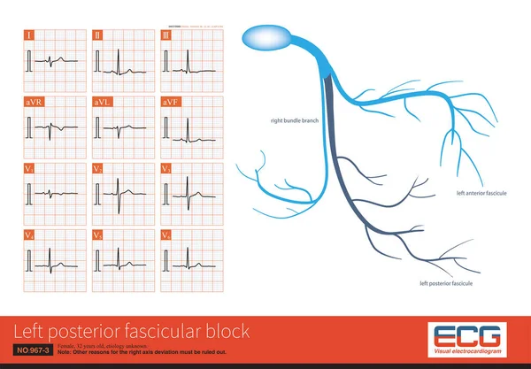
Stock image Male, 48 years old, paroxysmal palpitations for 10 years. ECG suggests ventricular preexcitation, and the bypass tract may be located in the left posterior septum.

Published: Jul.13, 2023 13:43:55
Author: asia11m
Views: 5
Downloads: 0
File type: image / jpg
File size: 13.19 MB
Orginal size: 10000 x 5781 px
Available sizes:
Level: beginner