Home
Images
Vectors
Videos
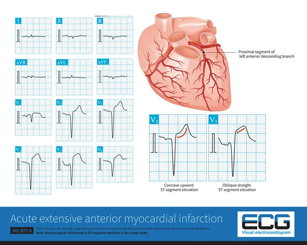
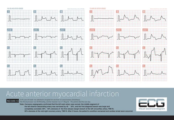
Stock image Male, 65 years old, admitted to hospital with chest pain for 2 hours. ECG showed hyperacute T waves. Coronary angiography showed occlusion of the middle segment of left anterior descending artery.
Published:
Sep.04, 2023 07:37:46
Author:
asia11m
Views:
5
Downloads:
0
File type:
image
/
jpg
File size:
14.03 MB
Orginal size:
10000
x
6620
px
Available sizes:
S
(500 x 331)
M
(1000 x 662)
L
(2000 x 1324)
XL
(10000 x 6620)
EL0
(10000 x 6620)
Level:
beginner
Download Image
Image topics:
Graphic
Illustration
Human
Healthy
Medicine
Medical
Line
Cardiology
Heart
Pulse
Body
Disease
Education
Science
Death
Organ
Lead
Graph
Rhythm
Diagnosis
Heartbeat
Anatomy
Cardiogram
Cardiac
Ekg
Electrocardiogram
Atrium
Ecg
Anatomical
Myocardium
Heart Rate
Heart Disease
Chest Pain
Cardiac Arrest
Coronary Artery
Left Ventricle
Right Ventricle
Left Atrium
Sudden Death
Sinus Rhythm
Coronary Angiography
Acute Coronary Syndrome
ECG Monitoring
Ecg Education
Qrs Wave
Left Anterior Descending Artery
Culprit Vessel
T Wave Changes
Similar stock images
Male, 64 Years Old, Admitted With Chest Pain For 4 Hours. The Clinical Diagnosis Was Extensive Anterior And Inferior Myocardial Infarction. The Culprit Vessel Was The Left Anterior Descending Branch.
10000 × 7987
Male, 75 Years Old, Clinically Diagnosed As Acute Anterior Septal And High Lateral Myocardial Infarction.The 12 Lead ECG Showed A Large Inversion Of T Wave With Prolonged QT Interval.
10000 × 5636
Acute Left Main Artery Occlusion Can Cause Both ST Segment Elevation And Non ST Segment Elevation Myocardial Infarction, Regardless Of Which Type, The Risk Of Death Is High.
10000 × 7584
This Is An Electrocardiogram Of Left Ventricular Hypertrophy With Left Axis Deviation. The QRS Electrical Axis Of The Frontal Plane Is Located In The Upper Left Quadrant.
10000 × 9612
Female, 57 Years Old, Diagnosed With Mitral Stenosis 6 Years Ago. This Electrocardiogram Had An Atrial Premature Contraction, Indicating Its Origin In The Right Superior Pulmonary Vein.
10000 × 6733
Sometimes, Because The QRS Axis Is In The Upper Left Quadrant, The High-amplitude R Wave Of Left Ventricular Hypertrophy Occurs In The Limb Leads, And Left Chest Leads Is Normal.
10000 × 10111
When The Middle-distal Segments Of The LAD Are Occluded, The ECG Of Extensive Anterior Myocardial Infarction Will Also Be Generated, But The Infarction Range Is Less Than That Of High Occlusion.
10000 × 8439
A 69 Year Old Patient With Acute Myocardial Infarction, Whose Infarction Affected The Anterior Wall, High Lateral Wall, And Inferior Wall, Ultimately Died During Hospitalization.
10000 × 6873